

No—both are MRI-safe, but gold may affect X-ray clarity while diamonds usually don’t.
At Lema Dental Clinic in Türkiye, the scenario where extravagant aesthetic touches meet medical necessity is all too common. Perhaps you’ve transformed your smile simply by placing a gold crown or a diamond-encrusted design, and it looks fantastic. But suddenly a medical necessity arises – such as an MRI for a headache or a routine dental X-ray – and suddenly your investment becomes risky.
It is even beyond whether or not it is safe. Actually, it almost becomes a question “Will this mess up my diagnosis?”
From our dental experience, demystifying myths from material science is the key initial step in answering this question. Basically, we have to get rid of the confusion first.
Can an MRI Rip Out Gold Teeth? The Truth About Dental Metals
There is a scary scene in a movie where, the moment an MRI machine is switched on, metal pieces start flying in the room. It is perfectly normal for patients imagine that their gold teeth might go through the same thing.
Let me tell you what actually happened: Gold is na on-ferromagnetic material.
Gold is simply nowhere near being attracted magnetically as much as iron or steel. That is the reason why the gold filling or crown that you have in your mouth will not be pulled, twisted, or heated significantly when you are inside an MRI scanner. Professor Doctor Coşkun Yıldız often tells his patients that dental gold of high-grade—in fact, an alloy—retains its stability. Actually, this is the plus one gets when buying from a high-end dental material range as compared to the cheap, nickel-rich ones that are probably reactive or even get warm during an MRI.
Still, there is one more way that it turns out to be a misbehavior, although it won’t come flying at you.
“Starbust” Phenomenon
The star-like streaking artifact is a direct consequence of the MRI machine interacting with gold.
Think of an MRI scan as a completely quiet, calm pond. The surface is so smooth that you can tell that the sky is there. This is one of the cases where an MRI scan and the still-water analogy are a perfect match. Just like where the stone thrown into the pond causes the waves, gold disrupts the magnetic field. These waves result in an artifact called “susceptibility artifact” or “starburst” pattern which is the one radiologists are very familiar with. The picture is referred to as a black hole or a white smeared patch which can be hiding the local anatomy. E.g. a doctor may be struggling to see the most important details when diagnosing a patient’s brain or TMJ if the patient’s gold grill is so large that it blocks the view.
Why Traditional X-rays Could Not Work Around the Metal?
X-rays relate to the density of an object. They are capable of passing through soft tissues quite easily like gums but they get blocked when solid structures, such as bones are encountered. In this way the contrast is formed in the images.
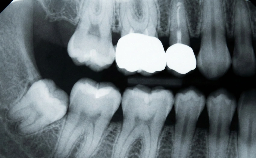
Due to the great density of gold, it acts as a solid lead wall that X-ray beams cannot penetrate rather than a window. This is the sort of stuff Dentist Polen Akkılıç and her dental team in Turkey face as part of their daily routine. The X-rays’ radiation can’t go past the gold compounds, and that’s why the use of conventional 2D radiography is not possible to see under a crown or a grill.
In case there is a cavity which started forming beneath a gold crown, the X-ray will reveal the spot only when it is already quite large and has reached the edges. Therefore, tactile examination is given more emphasis and sometimes we resort to 3D imaging (CBCT) so that we can get a nice clear view of the metal “shadow” surroundings.
What are Diamonds Made of Then?
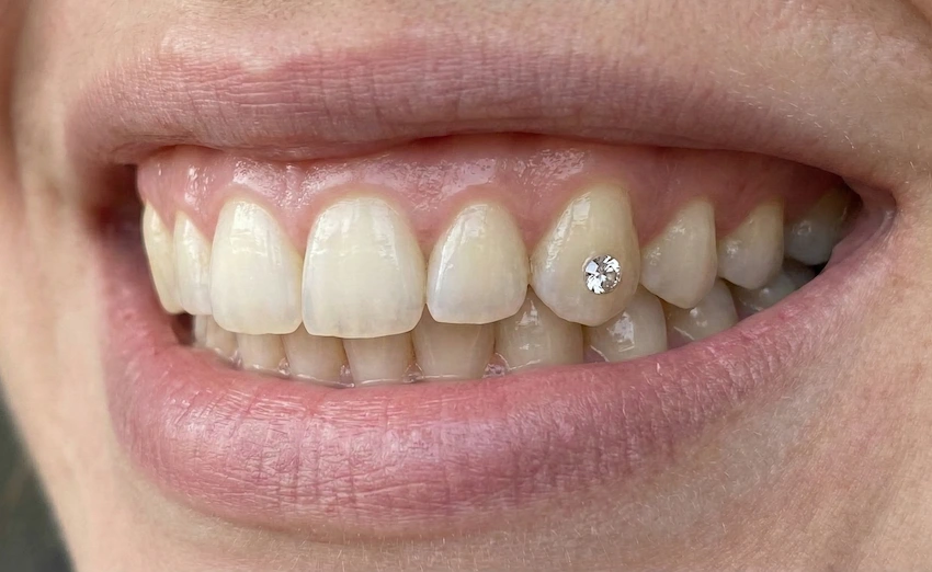
Diamonds are basically pure carbon. Interestingly enough, a pure diamond is slightly radiolucent; in other words, X-rays penetrate it even more than dental enamel.
There is a catch though: Hardly ever do you attach a diamond directly to the tooth with no metal backing. Usually, it is the backing metal in the setting—most frequently gold or platinum—that causes the X-ray or CT scan scattering. The jewel is perfect; it is the metal setting that complicates the matter.
Material Differences: Matching the Choice of Your Smile
Lema Dental Clinic’s dental team during a smile makeover consult will not simply discuss the dental materials with you but also connect those materials to your way of life.
| Material | Magnetic (MRI Risk?) | X-Ray Visibility | Artifact / Blurring Risk |
| High-Carat Gold | No (Safe) | Blocks radiation (radiopaque) | Moderate to high |
| Diamonds | No (Safe) | Low visibility (radiolucent) | None (stone itself) |
| Amalgam (Silver) | No (Safe) | Blocks radiation | Moderate |
| Zirconia | No (Safe) | Similar to bone | Low (better clarity) |
| Titanium | No (Safe) | Radiopaque | Low (biocompatible) |
| Our Clinical Approach | All dental materials should be MRI-safe | Preference for low-artifact materials | Diagnostic clarity comes first |
Let’s imagine that a radiologist would be performing an MRI of your head or neck and therefore needs to know whether, ahead of a certain point, you would be able to remove any gold teeth.
- Fixed veneers/crowns: You cannot remove them. In order to reduce the scatter effect, the technician will adjust the machine’s settings.
- Removable grillz: You need to remove them. Not because they are high magnetism but simply because they degrade the image quality without any necessity.
We often say that the overall work quality matters a great deal. In the bid to imitate gold, cheap alloys often contain ferromagnetic (i.e., iron or nickel) impurities. That’s actually where the danger lies. At Lema Dental Clinic, we look into the matter of our materials in detail for you to be absolutely safe at all times—whether you are on our operating chair or inside the MRI tube.
Q&A: The Doctor Replies
Metal loops could theoretically transfer heat as a result of radiofrequency pulses, but dental gold merely makes this scenario a near-zero probability, and even then mostly as a barely palpable phenomenon. You may experience a slight sensation of warming but it will not be enough to give you a burning sensation.
The direct way to have it revealed is definitely not. Gold is completely immune to the X-ray. What we mainly do is monitor the edges of the crown and will the symptoms to confirm that there is decay under the crown.
In my opinion, almost certainly “no”. The diamond is pretty small and if the setting used is of a non-magnetic nature (such as gold), then no worries at all. However, in the case where the purpose of the scan is a precise focus on a very small spot right next to the diamond, any disturbance or artifact that results from the setting might be an issue. You are supposed to always inform your technician about this.
When it comes to strictly diagnostics, sure, it is. Zirconia is a metallic substance and therefore has the same strength as a metal but in relation to X-rays and MRI, it behaves far better. Gold, after all, doesn’t create giant “black hole” artifacts, while zirconia doesn’t.
Of course, you can. Nowadays, X-ray CT scanners utilize a metal artifact reduction (MAR) feature, which is basically a software helper. It is not a perfect thing—there will be some streaks still—but most of the time the doctor can see everything that is required.
- Behr, M., Hahnel, S., Faltermeier, A., Bürgers, R., Kolbeck, C., & Handel, G. (2010). The two-body wear of dental porcelain and metal-ceramic antagonists. Clinical Oral Investigations, 14(3), 291–302.
- Cruzeiro, M. M., & Melo, S. L. (2019). Influence of metal artifacts in magnetic resonance imaging of the head and neck region: A review. Journal of Oral and Maxillofacial Radiology, 7(1), 12-17.
- Eggers, G., Rieker, M., Welzel, T., & Mühling, J. (2005). Geometric accuracy of magnetic resonance imaging of the mandibular nerve. Dentomaxillofacial Radiology, 34(4), 225-231.
- Klinke, T., Daboul, A., Biffar, R., & Hirsch, C. (2012). Artifacts in magnetic resonance imaging caused by dental materials. PLOS ONE, 7(2), e31766.
- Shafiei, F., Memarpour, M., & Vossoughi, M. (2016). Effect of gold and amalgam restorations on the quality of MRI images. Journal of Dentistry, 17(2), 85-90.