
Surgery prevents pericoronitis, dentigerous cysts, and adjacent root resorption.
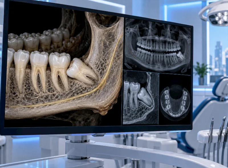
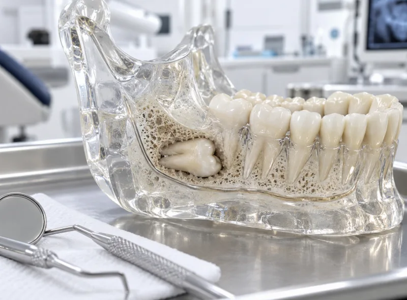
Impacted third molar is a kind of developmental disorder that occurs because of an anatomical conflict between the direction of eruption of the tooth, on one hand, and the space available in the corresponding jaw arch, on the other hand. If a wisdom tooth doesn’t have enough space to fully emerge through the mucous membrane of the gingiva, it stays either completely or partially buried in the alveolar bone. Although it is possible to wait in fully enclosed asymptomatic cases, when certain medical and biologic conditions occur, the necessity of preventive surgery becomes very clear.
The Pathogenesis of Pericoronitis
Pericoronitis is the main medical reason leading to extraction of the third molar. In the case of a partially impacted tooth, a flap of soft tissue over the erupting crown is called the operculum. Since such a pocket of periodontal space exists around a tooth that has been just partially impacted, there will also be a tendency for the soft tissues around the operculum to become infected, especially if food gets lodged between them and bacterial infection is allowed. This infection, which is usually caused by anaerobic bacteria, if not treated, becomes more severe, leading to pus formation, severe trismus (incapability to open the mouth), and may even spread and result in other infections of the deep fascial spaces of the face and neck area, for example, Ludwig’s angina, which may even cause death.
Odontogenic Cysts and Root Resorption
Not only the infection but also the long-standing effects of the third molar impaction are very destructive to the surrounding jaw area. In fact,
- Dentigerous Cysts: With every developing tooth, there is a surrounding sac called a dental follicle that later becomes a tooth root. Sometimes this follicular lining degenerates into an inflamed cyst filled with fluid. When the cyst grows it puts pressure to the area which, in turn, causes resorption of the healthy bone in the jaw that is close to the cyst and can result in a complete loss of the part of the mandible.
- External Root Resorption: A tooth that has a mesioangular eruption direction, i.e., a tooth growing with its root pointing towards the cheek (tilting forward), causes the tooth’s crown to constantly press the adjacent tooth’s distal root, which eventually becomes so severe that it is almost impossible to remove the latter without also getting rid of the former.
- Distal Cervical Caries: The portion of one tooth that is closest to another one, particularly of a non-erupted molar and a erupted molar, is very vulnerable and easily carious due to the difficult to reach spot being covered with the soft tissues and the hard tissues which can’t be cleaned or brushed. As a result, cariogenic bacterial plaque remains and it quickly causes severe, subgingival caries that result in destruction of the dentin.
Clinical Diagnostics and Surgical Protocol
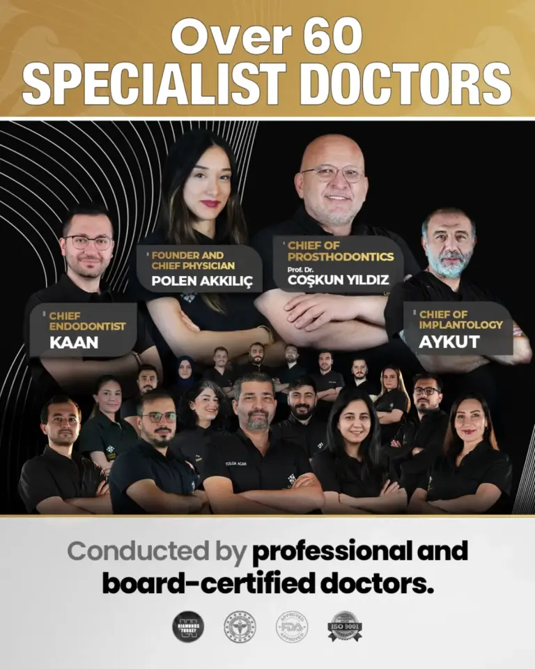
The determination of the exact location within space of the impacted molar at our dental center leads to defining the correct operating method of the surgeon. Our Dentist Polen Akkılıç, using 3D Cone Beam Computed Tomography (CBCT), is able to show with exactness the tooth’s proximity to the inferior alveolar nerve and the sinus floor of the maxilla.
The main operating method of Professor Doctor Coşkun Yıldız is completely atraumatic. First, he raises a mucoperiosteal flap, and then piezoelectric osteotomy, a technique that can preserve the overlying cortical bone is used to carefully remove the bone and afterwards the tooth is divided systematically into pieces. By doing so, the surgical trauma to the mandible is minimized and the surrounding alveolar structures are kept intact which is essential for hemostasis and proper bone regeneration.
Clinical Indications: Extraction vs. Observation
| Clinical Presentation | Maxillofacial Pathology | Mandated Treatment Protocol |
| Recurrent Pericoronitis | Acute anaerobic infection beneath the operculum. | Urgent surgical extraction to prevent systemic cellulitis. |
| Follicular Expansion > 3mm | Formation of a dentigerous cyst or ameloblastoma. | Enucleation of the cyst and surgical tooth extraction. |
| Mesioangular Pressure | External root resorption of the adjacent second molar. | Immediate extraction to salvage the healthy second molar. |
| Complete Bony Impaction (Asymptomatic) | Fully encased in alveolar bone with no cystic pathology. | Active clinical surveillance (annual panoramic radiographs). |
Frequently Asked Questions
1. Is it clinically possible for an impacted wisdom tooth to shift my other teeth?
Current orthodontic consensus indicates that third molars do not exert enough anterior vector force to cause primary crowding of the lower incisors. Late mandibular growth and physiological mesial drift are the primary biomechanical causes of late-onset crowding, not the wisdom teeth themselves.
2. Why do maxillofacial surgeons section the tooth during extraction?
Sectioning (odontotomy) is a conservative surgical technique. By using a specialized surgical bur to divide the crown and roots into separate pieces, the surgeon can remove the tooth through a much smaller window of bone. This drastically reduces post-operative edema, prevents jaw fractures, and protects the adjacent inferior alveolar nerve from crushing trauma.
3. What is paresthesia, and why is it a risk in lower jaw extractions?
The inferior alveolar nerve, which provides sensory innervation to the lower lip and chin, often runs directly adjacent to the roots of the lower third molars. If this nerve is bruised or stretched during surgery, it can result in paresthesia—a temporary (or rarely, permanent) numbness or tingling sensation in the lip and chin.
4. Can an impacted tooth be left alone if it doesn’t cause pain?
Lack of pain does not equate to an absence of pathology. Dentigerous cysts and external root resorption are typically asymptomatic until they have caused severe, irreversible damage to the jawbone or adjacent teeth. This is why routine 3D radiographic monitoring is a clinical necessity for all retained wisdom teeth.
Academic References
- Hupp, J. R., Ellis, E., & Tucker, M. R. (2013). Contemporary Oral and Maxillofacial Surgery (6th ed.). Mosby Elsevier.
- Mettes, T. D., Ghaeminia, H., Nienhuijs, M. E., Perry, J., van der Sanden, W. J., & Plasschaert, A. (2012). Surgical removal versus retention for the management of asymptomatic impacted wisdom teeth. Cochrane Database of Systematic Reviews, (6).
- Marciani, R. D. (2007). Third molar removal: an overview of indications, imaging, evaluation, and assessment of risk. Oral and Maxillofacial Surgery Clinics of North America, 19(1), 1-13.
- Kugelberg, C. F., Ahlström, U., Ericson, S., & Hugoson, A. (1991). Periodontal healing after impacted lower third molar surgery. A retrospective study. International Journal of Oral and Maxillofacial Surgery, 20(1), 18-24.