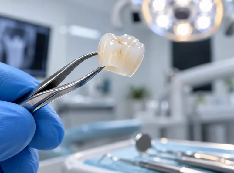

Replace crowns showing marginal microleakage, gingival inflammation, or material fracture.
Dental crowns are mechanical prostheses constantly facing a mixture of thermodynamic stress and heavy masticatory fatigue. With time, conventional restorations undergo structural degradation and chemical dissolution. The ability to diagnose the biomechanical failure of an elderly prosthesis in time clinically is critical to the prevention of irreversible necrotic changes in the dentinal and periodontal tissues underneath the prosthesis.
Prosthetic Failure Pathology Signs
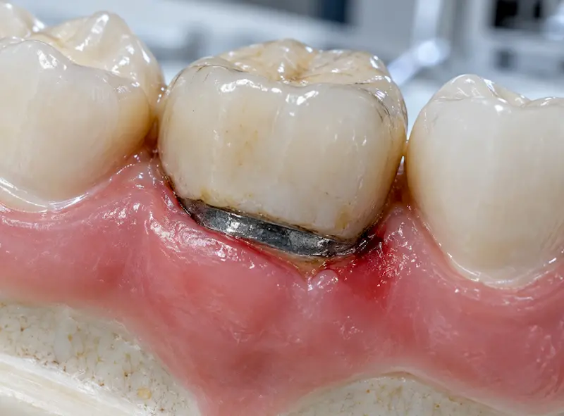
Prosthetic failure of dental restorations rarely happens immediately; they result in slow mechanical breakdown of dental tissue. A dental crown should be replaced if the following four clinical pathologies are present:
- 1. Microleakage along the border of the restoration: Any dental filling’s margin is going to be its weakest area. As the years go by, it is likely that the sealing cement which holds the restoration to the tooth starts wearing out. This small space created between the two allows cariogenic pathogens to enter the chamber and cause quick, undetectable development of secondary caries (decay) that is hidden under the prosthetic structure.
- 2. Violation of the Biological Width: The older or the inadequately shaped crowns are known to damage the gingival apparatus that anchors the gums. This constant physical disturbance leads to the periodontal diseases such as chronic localized periodontitis, marked by bleeding, pocketing and the loss of the supporting tissues, alveolar bone resorption.
- 3. Delamination (Separation) of Cohesive Materials: Traditional PFM (Porcelain-fused-to-metal) crowns have a two layer system which is weak and that is why the layers are likely to separate. Chewing is responsible for cyclic masticatory loading (chewing forces creating stress) that will result into development of microfractures in the veneered ceramic glass resulting to the shearing off of the surface from the metal substructure underneath.
- 4. Metal Oxidation of Metal Foundation (Dark Spot from an old filling in an adjacent tooth): The dark metallic neck of the crown is visible once periodontal tissues recede naturally with age. Moreover, base metal alloys oxidatively change the surface in the oral environment, releasing metal ions that leave permanent black/grey colored staining on nearby the gingival tissue.
Biomechanically, the Zirconia Treatment
Presently, evidence-based prosthodontics has come upon the use of Yttria-stabilized Tetragonal Zirconia Polycrystal (Y-TZP) as a mandatory dental material for upgrading an old restoration. The one single mass of a pure block of zirconium dioxide is what a monolithic Zirconia is made from resulting in the elimination of the likelihood of the interface being broken up or torn at the layers.
Upgrading at Lema Dental Clinic is a matter of strict digital precision. Dentist Polen Akkılıç, will be using an advanced intraoral scanner to record precisely the marginal shape of an abutment tooth.
This data gathering will then allow the lab to fabricate via CAD/CAM very bio-inert Zirconia crown that completely fits against the tooth margins so as to stop bacterial microleakage through. The Professor Doctor Coşkun Yıldız adds that the use of metal-free Zirconia does not only provide an increased bending strength (above 1,200 MPa) but it also eradicates entirely the galvanic activity which allows the gum tissue to grow back and tightly bind themselves to the shiny surface of the new ceramic implant.