

Yes—cavities can form under laminate veneers if the seal fails.
You decided after a long time deliberating. It is the truth. You are really desirous of that Hollywood smile through which some celebs and IG influencers flaunt their pearly whites. Nevertheless, at 2 a.m., your old doubting self sneaks in: What about my original teeth? Won’t they still be rotting right underneath that perfect white protective layer?
That can perfectly be explained. In fact, that is the very first question we get at Lema Dental Clinic in Turkey when somebody comes for a consultation.
The short answer? There is a possibility for cavities to develop under your laminate veneers.
Still, the truth is that there is more to it than what the short answer reveals. It is hardly ever the laminate’s fault. Most of the time, it is the failure of the seal, the hygiene neglect, or an incorrect initial placement. Let’s dump the promotional stuff for a moment and figure out how your smile’s biology functions as Professor Doctor Coşkun Yıldız would explain to you if you were his patient now.
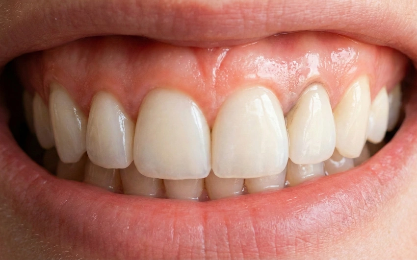
Do Laminate Veneers Really Protect Your Natural Teeth?

Porcelain is an inorganic material and therefore, similarly to glass and stone, it occurs naturally as bacteria-resistant. Bacteria cannot live on porcelain because there are no proteins and carbohydrates available for them. Hence, bacteria cannot infect or destroy porcelain in any way.
But the hero behind the dapping of the armor (your natural tooth) is still flesh and bone. If the armor has some defects, the arrow can find its way through it. In other words, it will be a flaw in the marginal integrity of the restoration.
Once the seal of your veneer is closed, your tooth will be less exposed to acids and sugars and thus even more over-protected. However, the minute the bond is broken, bacteria will enter the microspace between the porcelain and your tooth. Since this place cannot be cleaned with a toothbrush, the decay will gradually increase and continue expanding in size.
What Causes Cavities and How Can They Be Prevented?
Based on clinical experience we can say that cavities under veneers are formed due to the following reasons:
1. The “Marginal Gap.”
This is the thing that really matters. When a dental technician or a dentist leaves an edge or a gap at the margin between the veneer and the gum line, this becomes an ideal spot for plaque buildup. It is just like leaving the front door slightly open during a storm as the rain will eventually get in.
This is mainly why Dentist Polen Akkılıç and her team pay so much attention to the smallest details. Instead of going through the hassle of taking traditional impressions, we use digital technology which guarantees the accuracy of our scans down to the micron level. A perfect fit means bacteria do not stand a chance of entering the mouth.
2. Gum Recession
If you brush your teeth too hard repeatedly over time or have gum disease, your gums will recede. The gums getting pulled back will uncover the root of the tooth, which is not covered by enamel and also not protected by a veneer . An exposed root is very susceptible to decay because it is very soft.
3. Past History
If a dentist places a veneer on a partly decayed tooth without fully removing the decay, the dentist is basically sealing in the bacteria. Many times we see such cases as a part of the correctional work done in other clinics. At Lema, we take the problem from the bottom up. We certainly don’t put a house on a swamp foundation.
Common Symptoms and How to Identify the Problem
How can you tell that there might be an issue, as the porcelain is not transparent?
| Symptom | Indication | Priority |
| Sensitivity to hot or cold | Veneer bonding may be compromised, or decay may have reached the pulp | Intermediate – A dental consultation is needed soon |
| Dark discoloration on the gums | Imperfect sealing allows stained liquids to penetrate; whitening or resealing may be required | Very high – Visit a specialist as soon as possible |
| Rough feeling with the tongue | Veneer margin may be chipped or plaque may be accumulating in a small gap | Intermediate – A specialist should smooth or adjust it |
| Pain when biting | The tooth structure beneath the veneer may be cracked or fractured | Emergency – See your dentist immediately |
Lema Philosophy: Prevention at the Core of Dental Care
Maybe the main reason why you are visiting Turkey is to make sure the most skilled professional handles your case.
Professor Doctor Coşkun Yıldız points out that the survival of veneers depends (up to 80%) on the way they are carried out. In fact, we are very careful. Slowly and patiently trimming the enamel to lock in the veneer is akin to making a device that uses less glue.
Here are the ways we keep your natural teeth healthy when placing veneers:
- Above all, besides offering you the most comfortable and easiest treatment plan, we try not to remove that part of the enamel that can be left. This is because enamel is the main factor in the strength of adhesive cement for veneers. Therefore, the bond will be the strongest if the maximum enamel is preserved.
- We use only top-grade bonding agents that can almost entirely stop moisture penetration. Without that, no saliva (and, thus, bacteria) can get in between the tooth and the porcelain.
- The dental team is working under a microscope so that the patient can witness the gradual transition from porcelain to the tooth surface first-hand before leaving. One of their ongoing tasks is checking the margins.
Frequently Asked Questions (FAQs)
Regrettably, A violation of the answer is yes. First, we can only suspect decay under the porcelain, but we are not able to get at it with a drill without cutting or taking off the veneer. But after the removal of the veneer, the process is similar to the new veneer that is fabricated to cover the tooth which has just been prepared.
This is where you need to be careful. I generally advise my patients against using rough “whitening” toothpastes that contain baking soda or charcoal. These toothpastes per se are not tooth decay-causing agents but they definitely will scratch porcelain surfaces. These scratches although very small are prone to plaque aggregation which leads to an increase in bacteria in the mouth and causes compromise of veneer margins. Therefore, you should limit yourself to the use of only non-abrasive toothpaste gel.
It used to be so. However, nowadays, bonding resins and cements are so powerful that they would hardly be noticeable if there is a decrease in the cement even throughout the entire period. Unless some accidents happen such as opening a bottle with your teeth or some contamination during the bonding process that leads to bond breakage, the chemical bonding so formed is often stronger than the tooth itself.
Under no circumstances are you expected to attend a meeting with us in Istanbul every six months. On the contrary, if you think that visiting the dentist twice a year is beneficial for professional tooth cleaning (scaling), why don’t you just do that in your local area? Meanwhile, in case your bite is not satisfactory, you should immediately get in touch with us. We are able to utilize digital X-rays along with the provision of instructions to the local practitioner for the checks they need to carry out.
Ordinarily, you won’t. The problem is that the nerve is not stimulated through the veneer so one wouldn’t usually get the initial cavity pain. That being said, X-rays should be taken without fail. The only way to “see through” the armor is to check the “knight’s” health, that is the closest to the tooth under the veneer.
- Peumans, M., Van Meerbeek, B., Lambrechts, P., & Vanherle, G. (2000). Porcelain veneers: a review of the literature. Journal of Dentistry, 28(3), 163-177.
- Layton, D. M., & Walton, T. R. (2013). An up to 16-year prospective study of 304 porcelain veneers. The International Journal of Prosthodontics, 26(4), 336-358.
- Friedman, M. J. (1998). A 15-year review of porcelain veneer failure—a clinician’s observations. Compendium of Continuing Education in Dentistry, 19(6), 625-630.
- Magne, P., & Belser, U. (2002). Bonded Porcelain Restorations in the Anterior Dentition: A Biomimetic Approach. Quintessence Publishing Company.
- El-Mowafy, O., & Wang, J. F. (2009). Longevity and clinical performance of IPS-Empress ceramic restorations—a literature review. Journal of the Canadian Dental Association, 75(8), 579-581.