

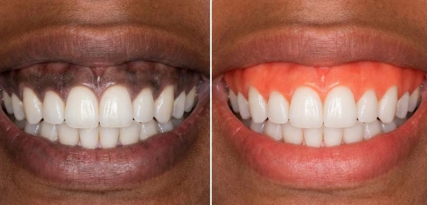
Pink gums in one visit.
It is quite possible that you have spent a considerable amount of time worrying about the whiteness of your teeth. There are many things you could have thought of doing, such as veneers, whitening strips, or alignment.
However, if we take a step back and check the “frame” of your smile, we will see that it’s not all about the enamel. A gorgeous painting can even lose its charm if it is put in a dark, heavy frame, bright white teeth can look harsh and unnatural if surrounded by dark, pigmented gums.
In medical terms, this phenomenon goes by the name of gingival hyperpigmentation. During our work at Lema Dental Clinic, we notice that it is almost a daily occurrence when a patient, who seems very confused, explains that they came to Turkey for a holiday and ended up with a dental treatment. The major reason for the visit was that the patient would always smile and love their teeth, but they would still hide the smile.
The problem is not enamel but simply melanin. Dark gums, no matter what the reason for them is, such as genetics, smoking, or medication, give an unbalanced and unhealthy look to a set of teeth even when the teeth are totally healthy and disease-free. However, it’s in a single appointment and most likely within forty-five minutes that we can restore your gums’ color to a natural coral pink.
Why Do Gums Turn Dark?
Melanocytes are cells that skin has, which make melanin, which protects your skin from the sun. Your gums also have those cells.
Professor Doctor Coşkun Yıldız regularly uses the analogy of how the skin gets covered by a “deep tan” that never goes off, which is a hint for the patients to understand that the same thing – gum pigmentation – is a form of sun tanning.
In the case of our international clientele, the dark color is just the result of anatomical variation. This is simply the result of the tobacco chemicals stimulating these cells to be more active and thus producing the formation of brown or black spots on the gum tissue; nonetheless, there is a particular condition called “Smoker’s Melanosis.”
So how is it possible to get rid of the color without damaging the tissue?
The Laser Advantage: Vaporizing, Not Cutting
At the time of treatment with the scalpel only, the top layer of the gum was removed by very precisely slicing. After such surgeries, the patient has suffered a lot of pain and bleeding, and the recovery time has been quite long.
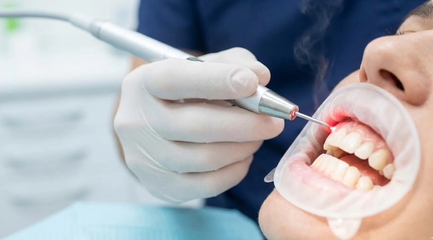
Nowadays, Dentist Polen Akkılıç and her team work with high-frequency diode lasers. Don’t picture the laser as a knife but as a smart eraser.
The light’s particular wavelength is attracted to melanin and water. The laser vaporizes the melanin-containing cells instantly, and at the same time, blood vessels are sealed after the laser tip passes along the gum.
This is how the clinic looks to us:
- No bleeding at all: The blood vessels are closed by the laser as it works.
- Local anesthesia may not be necessary: A powerful numbing gel is generally enough, although for complete comfort, we use a local anesthetic.
- The outcome is visible right away: You enter the clinic with dark gums and leave with the pink ones.
Causes of Gum Hyperpigmentation
Besides genetics which are by far the most influential factor, we also come across some other causes during our appointments:
- Physiological (Genetic): It occurs normally in people with darker skin pigmentation; they are absolutely healthy but some of them may not like their appearance.
- Smoker’s Melanosis: The mixed chemicals in the tobacco bind to melanin and the lower and upper gums, especially in front, are mostly affected.
- Medication-Induced: The use of some antimalarial and antidepressant drugs may cause changes in pigmentation.
- Amalgam Tattoos: The grey/blue spots are formed due to small particles from silver fillings which have been trapped in the gum tissue.
Traditional Surgery vs. Laser Therapy
For your greater understanding of our rationale behind giving the laser treatment an exclusive priority at Lema Dental Clinic, we have laid out the differences between the old surgical method and our modern protocol.
| Feature | Surgical Peeling (Scalpel) | Lema Laser Depigmentation |
| Precision | Low (Hard to control depth). | High (Micron-level control). |
| Bleeding | Moderate to Heavy. | None to Minimal. |
| Pain Level | Moderate (Requires painkillers). | Mild (often just a “tingle”). |
| Bandages | Periodontal pack (gum putty) required. | No bandages needed. |
| Healing Time | 7–14 Days. | 2–4 Days. |
| Risk of Infection | Moderate (Open wound). | Very Low (Laser sterilizes). |
Clinical Answers on Gum Depigmentation
For genetic pigmentation, the results are incredibly long-lasting. In many cases, one treatment lasts a lifetime. However, “Smoker’s Melanosis” is different. If you continue to smoke after the procedure, the melanin will eventually return because the chemical trigger is still present. We view this treatment as a fresh start—a perfect reason to quit.
Most patients describe the sensation as a warm vibration or a light “snap,” similar to a rubber band. Because Dentist Polen Akkılıç uses local anesthesia, you feel nothing during the actual procedure. Afterward, the gums might feel slightly raw, like you burned them on hot pizza, but this sensation typically fades within 24 hours.
Yes, but with caution. Since your gums will be tender, we recommend avoiding spicy, acidic, or very hot foods for roughly 48 hours. Stick to a “soft white diet”—pasta, yogurt, fish, and eggs—while the tissue heals.
Immediately after the laser treatment, the gums will appear white (from the fibrin layer that forms to protect the tissue). This is a natural bandage. Over the next 2-3 days, this white layer sheds, revealing the fresh, pink tissue underneath. It is not a “scary” healing process.
This is where the expertise of the clinician is vital. Laser settings must be adjusted based on the biotype (thickness) of the patient’s gums. In Turkey, we utilize advanced diagnostic imaging to ensure we only target the epithelial layer where the pigment lives, leaving the underlying connective tissue untouched.
Kathariya, R., & Pradeep, A. R. (2011). Split mouth de-epithelization techniques for gingival depigmentation: A case series and review of literature. Journal of Indian Society of Periodontology, 15(2), 161-168.
Yousuf, A., Hossain, M., & Crowe, T. (2020). Clinical efficacy of diode laser in gingival depigmentation: A systematic review. Journal of Lasers in Medical Sciences, 11(3), 335-341.
Ribas, M. A., et al. (2019). Gingival melanin depigmentation with diode laser: A clinical case report. Journal of Cosmetic Dentistry, 35(1), 58-64.
Giannelli, M., et al. (2018). Comparative evaluation of diode laser and scalpel technique for gingival depigmentation: A randomized clinical trial. Lasers in Medical Science, 33, 1617-1623.
Dummett, C. O. (1998). Oral pigmentation: Physiologic and pathologic. New York State Dental Journal, 64(4), 26-30.