

Pterygoid implants avoid sinus lift surgery and allow faster tooth replacement.
You are lying on the dental chair, thinking that your doctor will come up with a simple solution to replace your upper teeth. However, the doctor then points to your X-ray and utters the words that you dread: “You do not have enough bone. We have to do a sinus lift.”
The truth is, the problem of severe bone loss in the upper jaw presents a very common complicating factor. For a long time, the conventional treatment consisted of performing a sinus lift—a procedure in which the surgeon packs bone graft material under the thin sinus membrane (approximately as thin as the skin inside an eggshell) and then waits for 6 months to complete the healing process. Only after that could implants be placed.
However, let’s examine the details a bit. Suppose you did not have to do bone grafting at all? What if you could leave the clinic with fixed implants in just one day?
From our point of view at Lema Dental Clinic in Turkey, the goal of advanced implantology is to inflict the least possible trauma while producing maximum results. It is exactly from there that pterygoid (wing) implants start their story.
What Are Pterygoid Implants?
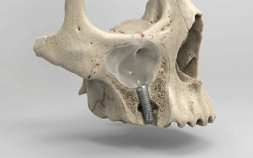
Imagine your upper jaw as the floor of a house. If the front yard soil is too loose or has been eroded for years, you don’t have to bring new soil and wait for it to settle necessarily. Instead, you can drive longer and stronger piles deep down to the original, solid bedrock at the rear of the property.
Pterygoid implants are unusually long dental implants, generally measuring between 15 mm and 20 mm. Instead of being positioned vertically into the delicate, hollow bone underneath the sinus cavity, the implants are inclined very sharply backward. They get their fixation in the very pterygomaxillary region—which is a thick, very stable column of bone at the point where the upper jaw fuses with the base of the skull—directly.
As this deep bone never undergoes resorption or shrinkage, even after several decades of being toothless, it offers a very firm base.
The Clinical Reality: Why We Choose the “Wing” Approach
Professor Doctor Coşkun Yıldız often points out that the most important factors in the total change of the treatment which would have otherwise been long and painful are patient comfort, and treatment duration. When we avoid the sinus, we avoid the need for an invasive graft. We get rid of the painful six-month healing time.
This is what we keep on repeating in our clinic. Patients here who were told they were “uuntreatable become locals with fixed, functional teeth in a few days after flying to Turkey, having undergone surgery.
Pterygoid Implants vs. Traditional Sinus Lifts
Let’s put the features and benefits of the pterygoid implants and the traditional sinus lift side by side to grasp why this method has become a revolution in full-mouth rehabilitation.
| Feature | Traditional Sinus Lift & Implants | Pterygoid (Wing) Implants |
| Bone Grafting Required? | Yes. Often requires artificial or autogenous bone grafting. | No. Uses the patient’s existing bone structure. |
| Treatment Timeline | 6–9 months (sometimes up to 1 year). | Immediate loading often possible within a few days. |
| Anatomical Risk | Risk of perforating the sinus membrane. | Does not enter or pass through the sinus cavity. |
| Long-Term Success | Good, but depends on successful graft integration. | Very high success rate (over 95%) with stable fixation in dense bone. |
The issue still stands: Is it the right choice of treatment for everyone?
It requires a highly skilled surgical team capable of safely navigating the complex anatomy of the posterior maxilla. Dentist Polen Akkılıç and her team at Lema Dental Clinic meticulously plan every step with the aid of 3D CBCT imaging. Precision is key. The pterygoid plate is a deep and complicated structure, and the placement of implants there necessitates specialized, hands-on surgical training to which many small local clinics may not have access.
Frequently Asked Questions
Not really. We actually don’t cause trauma to the area and get the same result by using the patient’s natural bone structure as a scaffold which means that there are no sinus membrane lifts or bone graft packing. The majority of our patients have a smaller amount of swelling and thus recover more quickly. The whole procedure is done under local anesthesia or sedation, so basically you wouldn’t even know that it happened.
Usually, a single visit of about 5 to 7 days is enough for us to place the implants and attach your temporary fixed bridge. Almost immediately, without multiple international trips, you get your smile back.
Age is a factor very rarely considered that much; bone quality and general health are more important. Since these implants are fixed in a part of the skull that doesn’t get older, they are an ideal solution for elderly people who have been through severe bone loss over a long period of time.
No, you won’t be aware of them once they are healed. They are deep within the bone and perform the same functions as your natural tooth roots.
It boils down to specialized training. The pterygoid area is complex anatomically. A comprehensive knowledge of surgical anatomy and ample clinical experience are prerequisites. Most dentists receive training only in standard implants and basic sinus lifts, hence their default to that standard recommendation.
- Balshi, T. J., Wolfinger, G. J., Slauch, R. W., & Balshi, S. F. (2013). A retrospective analysis of 152 pterygomaxillary implants: A 10-year study. The International Journal of Oral & Maxillofacial Implants, 28(2), 584-589.
- Candel, E., Peñarrocha, D., Peñarrocha, M., & Peñarrocha, M. (2012). Rehabilitation of the atrophic posterior maxilla with pterygoid implants: a review. Journal of Oral Implantology, 38(S1), 461-466.
- Graves, S. L. (1994). The pterygoid plate implant: a solution for restoring the posterior maxilla. The International Journal of Periodontics & Restorative Dentistry, 14(6), 512-523.
- Rodríguez, X., Vela, X., Segura-Mori, L., & Pérez-López, J. (2016). Anatomy of the pterygomaxillary area for implant placement: a cone beam computed tomographic study. The International Journal of Oral & Maxillofacial Implants, 31(2), 374-378.
- Tulasne, J. F. (1989). Implant treatment of missing posterior dentition. In The Branemark Osseointegrated Implant (pp. 417-426). Quintessence Publishing.