

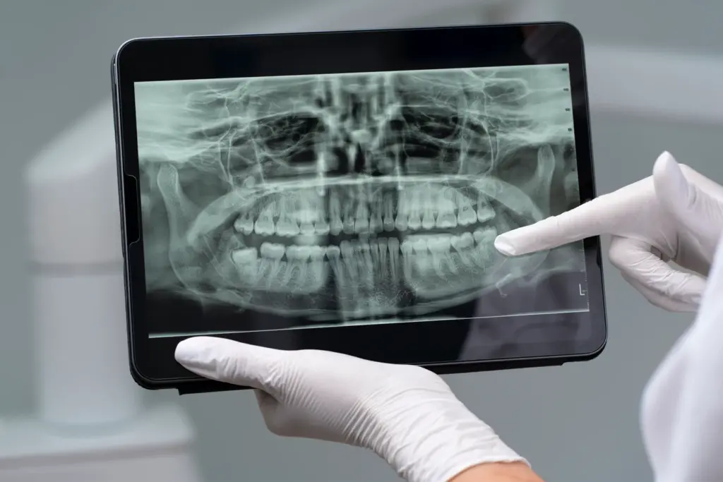
A panoramic X-ray shows your whole jaw to detect hidden dental problems.
Think of it this way; trying to restore a heritage building without even once consulting the blueprints is pretty much the same as one might repaint the walls and repair the windows without knowing if the foundation is crumbling or if the wires behind the drywall are prone to cause a fire.
To a dental surgeon, it similarly feels like performing a treatment on a patient without a Panoramic X-ray (OPG).
Looking Beneath the Surface
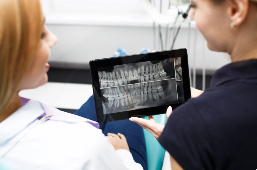
While a typical intraoral X-ray (the small one that you bite on) is very suitable for identifying tooth decay between the teeth, it is just like peering at a room through a keyhole; you get to see the details but the whole picture is lost.
By contrast, a panoramic X-ray is like a camera with a wide-angle lens. It can reveal the entire oral structure in a single photo: the upper and lower jaws, the temporomandibular joints (TMJ), the nasal sinuses, as well as the mandibular nerve canal.
For instance, a professor, Doctor Coşkun Yıldız, likens this diagnostic measure to a pilot who is inspecting the weather radar before the flight. You may be able to take off without it, but what you are doing is seriously compromising your safety. In Turkey, where many international patients come to us for full mouth rehabilitations, it simply is not an option to operate without this information.
What We Often See Hidden
Besides cavities, when Dentist Polen Akkılıç and her team assess a panoramic scan, they look for various other things that may not be apparent but could negatively affect your health or drain your wallet.
Our regular discoveries are:
- Besides being ticking time bombs, they can be pressing the roots of healthy molars or have cysts in them.
- If you are thinking about getting implants, we will find out whether your “soil” is deep enough to hold the “tree.”
- Some patients have root tip (apical periodontitis) infections that develop silently over several years and gradually destroy the jawbone, without the patient ever experiencing pain.
- When we are placing upper implants, we have to know where the sinus floor is so that we’re not going to perforate it.
The Safety Question: Radiation or Risk
Radiation exposure is frequently mentioned as one of the reasons against the use of X-rays. It is essential to put the issue into perspective. Technology in Turkey has reached a level of rapid advancement. Digital panoramic devices now emit such a small amount of radiation compared to the film-based ones that the latter are no longer used.
If we speak very accurately, a single panoramic X-ray provides a radiation exposure of approximately 0.01 mSv. As a matter of fact, for the sake of comparison, the cosmic radiation during a cross-country flight alone will expose you to a dose of about 0.04 mSv at high altitudes. In fact, the radiation dose you will receive on the plane flight to Istanbul for your dental treatment is more than the one from the diagnostic scan.
Diagnostic Comparison: Which Scan Do You Need?
Radiographs are not interchangeable. Different “weapons” are employed based on the clinical need.
| Details | Periapical (Bitewing) X-Ray | Panoramic (OPG) X-Ray | 3D Tomography (CBCT) |
| Main Function | Detects tooth decay and single-root abnormalities | Provides full jaw overview, impacted teeth, cysts, structural issues | Guides implant placement, evaluates bone width, traces nerves |
| Level of Detail | Highly detailed, small area (1–3 teeth) | Moderately detailed, entire mouth | Ultra-detailed, three-dimensional view |
| Radiation | Very low (<0.005 mSv) | Low (0.01–0.02 mSv) | Moderate (depends on field of view) |
| Purpose | Annual check-ups | First visit / treatment planning | Complex surgeries / implant planning |
Lema Standard of Care
We are against ordering X-rays simply for the sake of it. We get them done only when Professor Doctor Coşkun Yıldız insists on being completely sure about your health situation. If we decide that veneers are the right solution, we have to be absolutely certain that the roots are healthy. If the implant option is chosen, then the mandibular nerve should be traced to prevent the risk of nerve damage (numbness).
Disregarding this measure would be akin to a skyscraper being erected on a swamp; it might appear sumptuous on the inauguration day, but the failure of the entire structure is a certainty. At Lema Dental Clinic, we aim for our constructions to last.
FAQ
It really depends. If they are digital, clear and recent (not older than 6 months), we can usually make use of them. However, if you have experienced pain or sensitivity, had a treatment done after the X-rays, or if the quality of the images is not good, a new scan will be necessary so that we can be certain no new issue is missed.
It may show large cavities, but it has very low sensitivity in detecting small “interproximal” decay which is the decay between the teeth. Thus, we generally supplement the OPG with a clinical examination and bitewing X-rays so that no part of the diagnosis is missing.
Usually, non-emergency X-rays are not done especially during the first trimester. However, in a dental emergency case (a severe infection, for example), the infection risk to the baby is often higher than the risk from an X-ray. In case the scan is the only option, a lead apron and a thyroid collar will be used for the utmost protection.
The entire process duration is negligible. You will be requested to simply stand still in the machine, bite on a plastic piece that is held by your teeth and the arm of the machine will revolve around your head. The whole time of exposure will be 14 to 20 seconds maximum. Also, the treatment, i.e., X-ray, is absolutely pain-free and none of the in-mouth parts is so deep as to cause discomfort to your sensitive gag reflex!
It’s a big misunderstanding! Essentially, veneers are only placed on healthy and sound tooth surfaces. When veneers are put on teeth that have root infections beneath or gum disease (which can be seen as bone loss on the OPG), the veneers won’t last long and will fail. First of all, we identify the problem and then we do the treatment to make it healthy. Next, we change the look.
- White, S. C., & Pharoah, M. J. (2014). Oral Radiology: Principles and Interpretation. Elsevier Health Sciences.
- Rushton, V. E., & Horner, K. (1996). The use of panoramic radiology in dental practice. Journal of Dentistry, 24(3), 185-201.
- Vandenberghe, B., Jacobs, R., & Bosmans, H. (2010). Modern dental imaging: a review of the current technology and clinical applications in dental practice. European Radiology, 20(11), 2637-2655.
- American Dental Association Council on Scientific Affairs. (2012). Dental Radiographic Examinations: Recommendations for Patient Selection and Limiting Radiation Exposure. ADA.
- Perschbacher, S. (2012). Interpretation of panoramic radiographs. Australian Dental Journal, 57(s1), 40-45.