

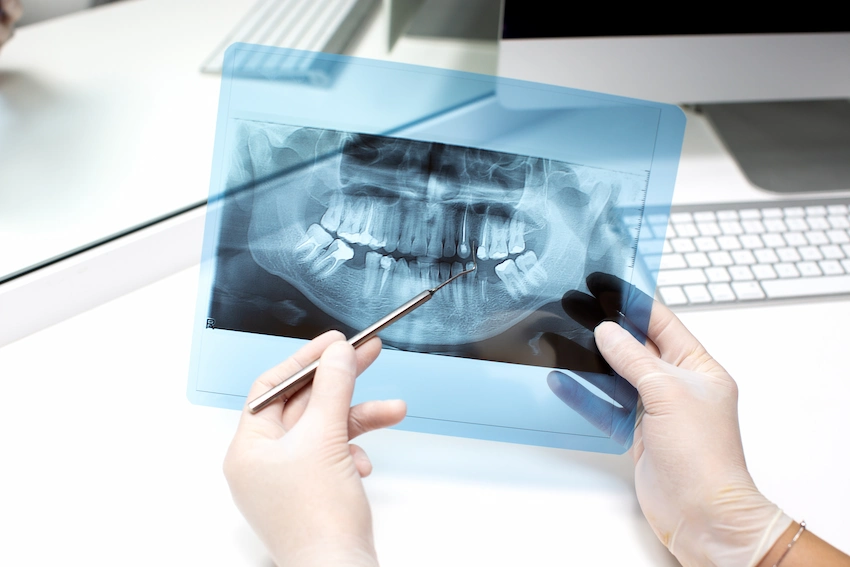
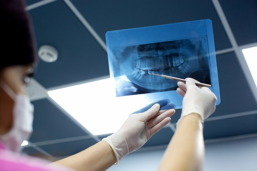
Dental X-rays reveal hidden details for safer surgery.
Imagine hiring an architect to build a luxury high-rise, but no matter how many times you ask, the architect refuses to check the soil before pouring concrete.
Surely, you wouldn’t give them such a project, would you
The fact is, the dental surgery is just the same.
At Lema Dental Clinic, Turkey, we consider 3D advanced imaging a very important part of your safety. It is not an additional step or an extra step. It is the most important step. Whether we plan a single implant or a full smile makeover, the decision about your dental surgery depends on what we cannot see with the naked eye.
Seeing the Unseen: Beyond the Gums
Your visible gums only tell the story when you are seated in the chair. The jawbone is the strong base of your face. If that base is weak, thin, or hollow, a titanium implant could be completely misplaced, probably leading to failure.
Professor Doctor Coşkun Yıldız always says that a surgeon’s hands cannot be more precise than the imaging guiding them. A great surgeon does not just make a guess. They map, measure, and then execute.
Navigating Hidden Hazards: Nerves and Sinuses
However, when we talk about the hidden map inside your mouth, the human jaw conceals very important structures. For example, a major nerve is just millimeters away from where a lower implant must be placed. This nerve is the one that conveys the feeling to your lower lip and chin.
Similarly, the risks are very high for the upper jaw too. This is because the sinus cavity is right above your back teeth. Dentist Polen Akkılıç and her team always remind our patients of one important fact. The sinus membrane is as thin and fragile as the skin inside an eggshell. Injuring it during surgery can lead to several problems.
We often observe that many of our patients come to us with very complex bone problems. And without a perfect 3D map, working around such delicate areas is simply not safe.
Old Tech vs. New Tech: Why We Choose 3D
Is it true that all X-rays are the same?
The answer is just a simple No. Flat standard X-rays may be okay for discovering a cavity. But in terms of the complex surgery planning, they are completely worthless.
Here is a simple explanation of why top surgical clinics insist on 3D imaging:
| Feature | Standard 2D X-Ray | 3D CBCT Scan |
| View | Flat, two-dimensional | Full 3D digital model of your jaw |
| Accuracy | Possible to be distorted | 100% exact millimeter precision |
| Bone Density | Almost impossible to tell | Displays exact bone thickness and health |
| Nerve Safety | Only a rough guess | Precisely maps the path of your nerves |
| Best For | Basic checkups | Implants, bone grafts, and surgery |
Frequently Asked Questions
Really, you are very safe. The Lema Dental Clinic scanners produce a highly focused beam of radiation. Which means that your exposure is extremely low. Besides, you are actually exposed to more background radiation during the flight to Turkey than from our quick digital scan.
Well, it is because bones change over time. Bone after tooth loss shrinks very fast. Using a flat old image is like using a ten-year-old map for driving in modern Istanbul. Today, we have to see exactly how your bone looks.
Not at all. The scan only lasts for about 20 seconds. You just have to stand still. The machine travels around your head. No pain at all, and there is instant, life-saving data given to us.
A single implant, even the one, always requires a perfect placement. We have to ensure that it is able to withstand the chewing pressure without damaging other roots or nerves. Precision is a must.
All guessing is out of the way. Even surgery is done virtually on our computers first before you sit in the surgery chair. You could be done with the exact implant size and angle. And if you need a bone graft, you know it immediately. Surgery will become faster, safer, and highly predictable.
- Bornstein, M. M., Horner, K., & Jacobs, R. (2017). Use of cone beam computed tomography in implant dentistry: current concepts, indications and limitations for clinical practice and research. Periodontology 2000, 73(1), 51-72.
- Tyndall, D. A., Price, J. B., Tetradis, S., Ganz, S. D., Hildebolt, C., & Fasbinder, D. J. (2012). Position statement of the American Academy of Oral and Maxillofacial Radiology on selection criteria for the use of radiology in dental implantology with emphasis on cone beam computed tomography. Oral Surgery, Oral Medicine, Oral Pathology and Oral Radiology, 113(6), 817-826.
- Benavides, E., Rios, H. F., Ganz, S. D., An, C. H., Resnik, R., Rebaudi, A., … & MacNeill, S. R. (2012). Use of cone beam computed tomography in implant dentistry: the International Congress of Oral Implantologists consensus report. Implant Dentistry, 21(2), 78-86.
- Harris, D., Horner, K., Gröndahl, K., Jacobs, R., Helmrot, E., Benic, G. I., … & Herman, S. (2012). E.A.O. guidelines for the use of diagnostic imaging in implant dentistry 2011. A consensus workshop organized by the European Association for Osseointegration at the Medical University of Warsaw. Clinical Oral Implants Research, 23(11), 1243-1253.
- Jacobs, R., Salmon, B., Codari, M., Hassan, B., & Leite, A. F. (2018). Cone beam computed tomography in implant dentistry: what dental educators need to know. Journal of Dental Education, 82(4), 434-443.