

White spots, brown shadows, holes.
Often, the first sign is a moment of panic in the bathroom mirror. You accidentally land with your floss, you bet a bit closer to you and suddenly there it is—a tiny, suspicious shadow on your molar. It is your imagination or what you see is a bit of coffee stain? A poppy seed? Or it is just the beginning of a root canal?
You feel troubled because tooth decay (caries) is a puzzle box. It doesn’t always have such a clear and distinct black color as the simple ones we see in pictures.
At our Lema Dental Clinic, we often encounter this situation where patients just don’t recognize early symptoms of the disease because they expect cavities to be holes in the teeth, whereas the disease actually starts most of the time very quietly. Professor Doctor Coşkun Yıldız can pinpoint tooth decay with an iceberg: the surface enamel that we see is a very small part of the total amount of decay underneath it.
One thing is for certain: it’s rookie moves to get your tooth crowned (and your credit card charged) when all you needed to do was to stop the cavity from forming. To do this, you first need to understand the eerie signs a tooth gives you before the hole makes its debut.
The Chameleon Effect: Stages of Visual Decay
Decay can be considered more as a gradual process than an instantaneous event. That is why the appearance of a cavity changes according to the depth level of the infection.
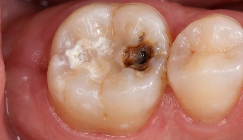
1. The “Chalky” Warning (Initial Demineralization)
Before the tooth decay process has reached a stage where the tooth surface has turned black, there is actually an emission of white color. Patient neglect is one of the main reasons for this stage, which is often overlooked. Maybe a spot near the gum line that is pale and has a bit of a matte finish is what you see. It could be mistaken for a very thin chalk piece or powder from a drywall dust container.
- The biological process: Nowadays, it is known that bacteria secrete substances called acids, which dissolve minerals such as calcium from not only the enamel but also the dentin. In this case, the outermost coating is still unbroken; however, it is becoming more and more brittle and less attractive due to the loss of water content.
- Positive side: At this phase one can get back the lost minerals and thus recover from the carious lesion but only if a high fluoride treatment is applied diligently.
2. The Brown or Black Stain (Enamel Decay)
As there is a breakage in the case of that the white part cannot resist it anymore and soabsorbstaining from the foodstuff, which can either be in yellow, brown or black. The novelty character of the feature (unique to a cavity) is the fact that a discoloration caused by a coffee stain appears as a big patch while that of a cavity is just a little dot.
- To touch a cavity with a dental explorer if it is just a discoloration Dentist Polen Akkılıç will feel that the tool is moving smoothly. If it is an active lesion, the tool will “stick” or catch. The enamel feels soft, like leather or wet wood, rather than hard glass.
3. The Shadow Under the Surface (Dentin Decay)
One of the most confusing things is that you sometimes get an impression that a tooth is perfectly fine – enamel is smooth, complete and shiny. The only thing that tooth seems a little bit off is that the inner part or just under the enamel is maybe a little bit grey or “bruised”.
- Let us take the example of a mother who comes up with the idea of baking an apple pie. The skin of the fruit looks fresh but the brown and squishy flesh inside indicates the rotten. This is how the stained core can be visible in teeth where bacteria have already reached the dentin, which is quite an extensive and shadowy area. , and it is differentiated easily by its color, i.e., the dentin is normally a darker shade of brown which “shines” throughthe translucent enamel,” as Dr. Polen explained.
The Hidden Danger: Interproximal Caries
Not all cavities are visible in the mirror. The truth is that the most lethal ones are actually hiding in the spots where your toothbrush can’t get to: strictly between the teeth.
You won’t see these until they are massive. Instead, you might notice:
- Floss fraying: When you floss, your floss always gets shredded or breaks at the exact same point.
- Food traps: Stringy meat or vegetables always get stuck in the exact same spot.
- Sharpness: Your tongue finds a jagged edge that wasn’t there last week.
What we have witnessed is that by the time you notice a cavity between your teeth visually, it has already substantially damaged the tooth structure by around 30-40%. This is the reason why a digital panoramic X-ray is an absolute necessity for diagnostics that we cannot do without here in Turkey.
Stain vs. Decay: How to Tell the Difference
The thing is, stains caused by your lifestyle and the initial stages of caries are often pretty similar. So what we usually do is take a quick look at them, comparing them according to these criteria.
| Feature | Surface Stain (Extrinsic) | Active Decay (Caries) |
| Color | Dark brown, black, or yellow. | Chalky white, grey, or black. |
| Distribution | Often follows the gum line or covers multiple teeth. | Isolated to a pit, fissure, or contact point. |
| Texture | Smooth and hard. | Soft, sticky, or rough/cavitated. |
| Cleaning | Can be polished off by a hygienist. | Cannot be brushed away; requires drilling. |
| Sensation | Usually painless. | Sensitivity to sugar, cold, or pressure. |
The Lema Protocol: Beyond the Naked Eye
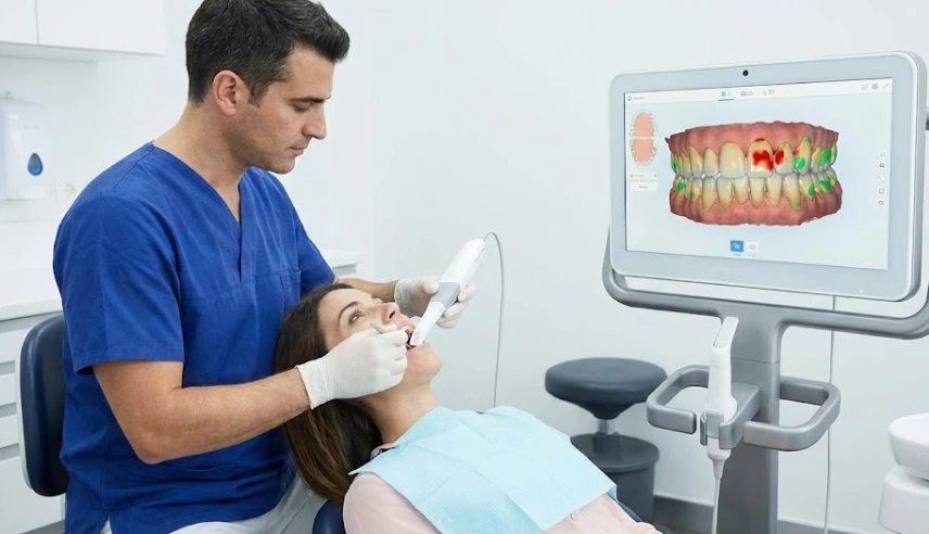
We do not rely solely on visual inspection. The human eye is limited. To ensure we aren’t drilling healthy teeth or missing hidden rot, Dentist Polen Akkılıç and her team utilize transillumination and intraoral cameras.
By shining a high-intensity light through the tooth, healthy enamel glows bright white. Decayed tissue, which blocks light, appears as a dark, distinct shadow. This allows us to map the exact boundaries of the decay before we even pick up a handpiece, ensuring we preserve the maximum amount of your natural tooth structure during your treatment in Turkey.
Top Queries on Identifying Tooth Decay
No, and this is a common misconception. Teeth have natural pits and fissures (grooves) on their biting surfaces. These grooves can trap stains that are impossible to brush out. If the black dot is hard, shiny, and pain-free, it is likely just a stain or “arrested decay” (an old cavity that stopped growing). We monitor these rather than drill them immediately.
A grey hue usually indicates internal issues. It could be deep decay casting a shadow through the enamel (as mentioned with the “rotten apple” analogy), or it could mean the tooth nerve has died due to previous trauma, causing the tooth to darken from the inside out. Both require immediate X-rays.
Surprisingly, yes. Active decay is essentially rotting organic tissue. It produces sulfur compounds that smell distinct—often described as a “meaty” or “rotten egg” odor that doesn’t go away after brushing. If you have chronic bad breath localized to one area of your mouth, it is a strong visual-olfactory sign of decay.
Professor Doctor Coşkun Yıldız warns patients: “Pain is a very late arrival to the party.” Enamel has no nerves. You will feel nothing while the decay is destroying the outer shell. You will only feel pain once the bacteria hit the dentin or the nerve pulp. If you wait for pain, you are usually waiting for a root canal.
On front teeth (incisors), decay often doesn’t look like a pit. It looks like a brown line hugging the gum tissue or a translucent/grey shadow on the edge where the teeth touch. In severe cases, a piece of the tooth edge may physically chip off because the structure underneath has crumbled.
- Fejerskov, O., & Nyvad, B. (2003). Diagnosis and classification of dental caries. Dental Caries: The Disease and its Clinical Management, 1, 1-8.
- Pitts, N. B. (2004). Modern concepts of caries measurement. Journal of Dental Research, 83(suppl 1), 43-47.
- Kidd, E. A., & Fejerskov, O. (2004). What constitutes dental caries? Histopathology of carious enamel and dentin related to the action of cariogenic biofilms. Journal of Dental Research, 83(suppl 1), 35-38.
- Ismail, A. I., et al. (2007). The International Caries Detection and Assessment System (ICDAS): an integrated system for measuring dental caries. Community Dentistry and Oral Epidemiology, 35(3), 170-178.
- Gomez, J. (2015). Detection and diagnosis of the early caries lesion. BMC Oral Health, 15(1), 1-8.