
Prevent cohesive fracture by managing bruxism and maintaining marginal seals.
Lithium disilicate (E-max) is a high-esthetic, glass-ceramic biomaterial frequently applied in modern prosthodontics because of its biomimetic optical properties and remarkable flexural strength (approximately 400 to 500 MPa). However, the longevity of these restorations in the mouth is not just a matter of the inherent strengths of the material itself. It depends on the micromechanical integrity of the adhesive bond and the management of occlusal (biting) forces. An understanding of the mechanism of ceramic degradation is essential for avoiding the occurrence of structural failure prematurely.
The Pathogeny of Ceramic Failure
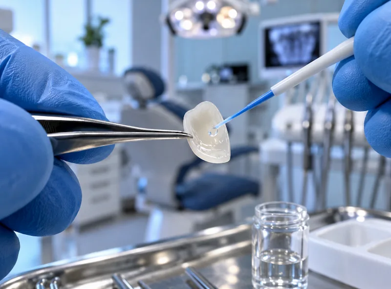
E-max veneers are extremely resistant when bonded to healthy enamel, but they are still affected by particular biomechanical and chemical factors during their lifetime. Structural failure generally occurs via one of three clinically observed modes:
- Cyclic Fatigue (Cohesive Fracture): Glass-ceramics are very sensitive to cyclic fatigue. For example, bruxism (nocturnal teeth grinding), a parafunctional habit, generates excessive shear forces which are not part of normal physiology. With time, such mechanical stress can be the cause of the development of microscopic cracks within the ceramic, leading to the final complete cohesive fracture of the veneer.
- Adhesive bond failure: It is through an intimate, complicated, chemical and micromechanical relationship that the veneer is retained. If the ceramic’s internal surface is not completely etched with hydrofluoric acid and treated with silane coupling agent, or the tooth substrate gets moist during cementation, then the resin cement will probably fail to adhere. As a consequence, the entire, undamaged veneer will fall off from the tooth.
- Marginal Microleakage: It is commonly found that marginal area of the restoration is the biomechanical weakness of any restoration. That is, the place where the ceramic and the tooth join. A microscopic gap is formed, once the cement is degraded by acidic environment. Such a space is exploited by cariogenic bacteria which initiate secondary demineralization of the enamel underlying the ceramic. In this way, structural integrity of the supporting abutment tooth is lost.
Largest Longevity by Clinical Procedures
At the Lema Dental Clinic, the survival of an E-max restoration through generations (decades!) relies first on the dentist’s chair time and also on patient’s commitment to cooperate.
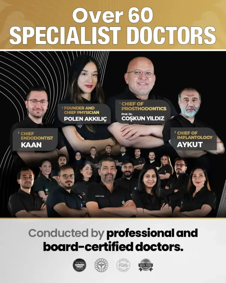
To build a micromechanically durable and impermeable seal of the cemented veneering units to the supporting tooth structure, Dentist Polen Akkılıç demands an almost perfect (98%) isolation by means of the rubber dam. The rubber dam serves as a barrier to saliva and crevicular fluid contaminating the bonding area, so that the hydrophobic resin cement can develop and maintain an optimal bond to the crystalline structure of the enamel, the hydroxyapatite.
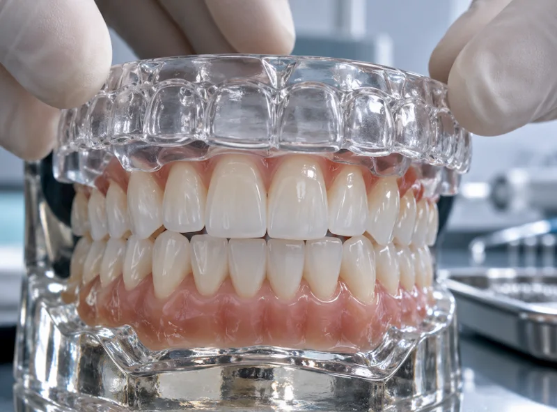
In order to reduce the threat of cyclic fatigue, Professor Doctor Coşkun Yıldız first examines his patient’s masseter muscle function as well as Temporomandibular Joint (TMJ) health. In cases where parafunctional biting (like presence of tooth grinding facets on posterior teeth) is the sign, he prescribes the patient to use a rigid acrylic occlusal splint (night guard) during sleep. This device redirects the damaging vectors of nocturnal bruxism, preserving the ceramic’s fragile integrity against the shear stress of sudden impacts.
Risk Factors and Clinical Interventions
| Biomechanical Risk Factor | Pathological Consequence | Clinical Preventive Intervention |
| Bruxism / Heavy Clenching | Cohesive fracture of the ceramic matrix. | Fabrication of a rigid acrylic occlusal splint. |
| Salivary Contamination | Adhesive failure and complete debonding. | Absolute rubber dam isolation during cementation. |
| Abrasive Prophylaxis | Destruction of the surface glaze. | Utilizing non-abrasive polishing pastes during hygiene visits. |
| Poor Oral Hygiene | Marginal microleakage and secondary caries. | Strict interdental cleaning and low-abrasive fluoridated dentifrice. |
Frequently Asked Questions
1. How long do Lithium Disilicate (E-max) veneers clinically survive?
When supported by precise adhesive bonding protocols and strict occlusal management, long-term clinical studies demonstrate a survival rate of 93% to 96% over a 10- to 15-year observation period.
2. Can an E-max veneer be repaired if it chips?
Minor incisal chips can occasionally be polished or repaired intraorally using a silane coupling agent and a highly filled composite resin. However, a major cohesive fracture requires the complete sectioning and replacement of the ceramic prosthetic to restore structural integrity.
3. Why must the tooth enamel be preserved during veneer preparation?
Enamel provides a vastly superior bonding substrate compared to underlying dentin. Bonding directly to the highly mineralized hydroxyapatite crystals of the enamel yields a predictable, high-megapascal bond strength that effectively prevents adhesive debonding and microleakage.
4. Do E-max veneers require a specialized toothpaste?
Yes. Highly abrasive dentifrices—such as whitening toothpastes containing coarse silica, baking soda, or activated charcoal—can mechanically scratch the polished surface glaze of the ceramic. This micro-abrasion leads to a permanent loss of optical translucency and an increase in bacterial plaque retention.
Academic References
- Guess, P. C., Schultheis, S., Bonfante, E. A., Coelho, P. G., Ferencz, J. L., & Silva, N. R. (2011). All-ceramic systems: laboratory and clinical performance. Dental Clinics of North America, 55(2), 333-352.
- Peumans, M., Van Meerbeek, B., Lambrechts, P., & Vanherle, G. (2000). Porcelain veneers: a review of the literature. Journal of Dentistry, 28(3), 163-177.
- Magne, P., & Belser, U. (2002). Bonded Porcelain Restorations in the Anterior Dentition: A Biomimetic Approach. Quintessence Publishing.
- Morimoto, S., Rebello de Sampaio, F. B., Braga, M. M., Sesma, N., & Özcan, M. (2016). Survival Rate of Resin and Ceramic Inlays, Onlays, and Overlays: A Systematic Review and Meta-analysis. Journal of Dental Research, 95(9), 985-994.