

3D CT Scans: The “GPS” of Modern Dental Surgery.
In the early days of dentistry, we relied on two-dimensional X-rays to see what was happening beneath the gums. While helpful, a 2D image of a 3D mouth is like looking at a shadow on a wall—it shows you the outline, but none of the depth. At Lema Dental Clinic, we believe that when it comes to your jawbone and nerves, “good enough” isn’t an option.
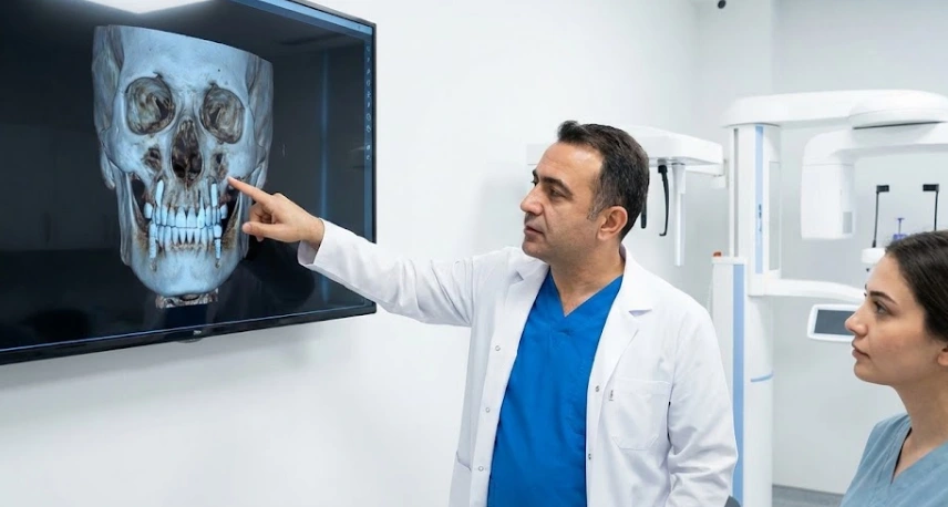
The reality is that dental surgery has moved into the realm of extreme precision. Whether you are getting a single implant or a full-mouth restoration, Professor Doctor Coşkun Yıldız often notes that a 3D CT (CBCT) scan is essentially our “Surgical GPS.” Without it, we are navigating your unique anatomy with a paper map; with it, we have a real-time, high-definition satellite view.
Seeing the “Invisible” Structures
Your jaw is a complex highway of nerves, blood vessels, and sinus cavities. The question remains: why can’t we just use a standard X-ray?
Standard X-rays suffer from “overlapping.” A nerve might look like it’s safely behind a tooth in a 2D picture, but in reality, it could be weaving right through the area where an implant needs to go. In our clinical experience in Turkey, using 3D imaging allows us to measure your bone density and volume down to the millimeter. This eliminates the “guesswork” and significantly reduces the risk of accidental nerve damage or sinus complications.
The Lema Protocol: Precision Means Faster Recovery
One thing Dentist Polen Akkılıç and her team emphasize to our international patients is that more technology actually leads to less surgery.
- Virtual Surgery: Before we even touch a scalpel, we perform your surgery on a computer screen. We “place” the virtual implant into your 3D bone model to find the densest, strongest anchor point.
- Flapless Procedures: Because we know exactly where the bone is, we can often perform “keyhole” surgeries that require fewer incisions and zero stitches.
- Predictable Outcomes: There are no “surprises” once the surgery begins. We know the height, width, and quality of your bone before you even sit in the chair.
2D Panorex vs. 3D CBCT Scan: The Comparison
| Feature | Standard 2D X-Ray | 3D CT (CBCT) Scan |
| View Type | Flat, overlapping image | $360^{\circ}$ volumetric view |
| Depth Perception | None (Shadow-based) | High (Millimeter-perfect) |
| Nerve Mapping | Estimated | Perfectly visualized |
| Bone Density | Cannot be measured | Detailed density analysis |
| Surgery Prep | General planning | Digital “Virtual” surgery |
FAQ: Insights from Professor Doctor Coşkun Yıldız
This is a common concern,” says Professor Doctor Coşkun Yıldız. “The truth is that modern CBCT scans used at Lema Dental Clinic use significantly less radiation than a medical CT scan you’d get at a hospital. It is a focused beam that only targets the jaw, making it a very safe tool for routine planning.
The number of teeth doesn’t change the complexity of the anatomy beneath them. Even for a single implant, we need to see the proximity of the neighboring roots and the thickness of the bone. In Turkey, we treat every case with the same level of diagnostic rigor.
Not at all. The scan itself takes about 10 to 20 seconds. You simply stand or sit still while the arm of the machine makes one quick rotation around your head. It’s completely non-invasive and open, so there is no feeling of being “trapped” like in a traditional MRI.
3D CT technology is a significant investment for a clinic. Many general practices still rely on 2D because of the overhead. At Lema Dental Clinic, we consider it an essential standard of care for any surgical procedure to ensure the highest success rates for our patients.
- Scarfe, W. C., & Angelopoulos, C. (2018). Maxillofacial Cone Beam Computed Tomography: Principles, Techniques and Clinical Applications. Springer.
- Jacobs, R., & Quirynen, M. (2014). Cavity or reality: the value of 3D imaging in implant dentistry. International Journal of Oral & Maxillofacial Implants.
- Bornstein, M. M., et al. (2014). Cone Beam Computed Tomography in Implant Dentistry: A Systematic Review Focusing on Guidelines and Protocols. International Journal of Oral & Maxillofacial Implants.
- Misch, C. E. (2007). Contemporary Implant Dentistry. Elsevier Health Sciences.
- White, S. C., & Pharoah, M. J. (2014). Oral Radiology: Principles and Interpretation. Elsevier.