

Yes, missing hidden dental infections.
You can sometimes feel like the request for a new set of X-rays is just a procedural formality when you sit in the dental chair for a routine check. Some patients hesitate, citing concerns about radiation or cost. However, in our clinical experience at Lema Dental Clinic in Turkey, we view X-rays not as an “extra” but as the primary scouting report for your oral health.
Skipping your dental X-rays is the same as telling your dentist to operate blindly. Of course, a physical examination is extremely important, but it only shows the doctor the surface of things. Below the gumline is a whole world of bone, nerves, and roots that cannot be seen without the help of instruments.
The “Sonar” of Dentistry: Seeing What Others Miss
Consider a dental X-ray to be the sonar of a submarine. It is unlikely that a ship captain would rely only on a pair of binoculars and still try to carefully navigate his ship through a dangerous coral reef. What he really needs is the knowledge of what is underneath the water. In the same way, Professor Doctor Coşkun Yıldız often points out that the most damaging dental problems, like interproximal caries or silent abscesses, can be without any symptoms for a long time and no one can expect their sudden deterioration till it is too late.
By the time you feel pain, the “small problem” has often evolved into an expensive, invasive emergency.
The Invisible Enemies: What We Find Beneath
- Interproximal Cavities: This type of decay is located between your teeth and is so difficult to detect that even a highly skilled dental explorer might not be able to find it.
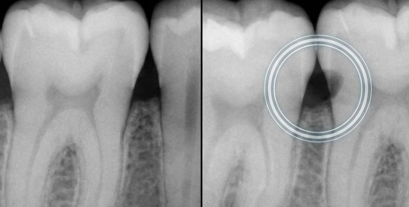
- Silent Root Infections: A periapical abscess may be present at the apex of the root and remain painless over many years while causing the gradual destruction of the adjacent bone.
- Wisdom Tooth Impaction: With the help of radiographs, we can monitor the development of wisdom teeth and see when they are about to hermetically press against second molars, similar to a slow-motion car crash happening underground.
Bone Loss: The Foundation’s Silent Erosion
One of the most significant risks of skipping X-rays is the inability to track bone density. We often compare the jawbone to the foundation of a building. If the soil underneath is washing away, the structure on top may appear stable until it collapses.
Dentist Polen Akkılıç and her team use digital radiographs to monitor the “crestal bone” levels. Gum disease (periodontitis) is a master of disguise; it often destroys the bone supporting your teeth long before your gums even appear red or swollen. Without an X-ray “map,” we cannot see if your foundation is being compromised.
Comparing the Diagnostic Power: Physical vs. Radiographic
The reality is that a physical-only exam is incomplete. The table below illustrates the critical gaps that skipping X-rays creates in your care plan.
| Diagnostic Tool | What It Detects | What It Misses |
| Physical Visual Exam | Internal decay, root abscesses, and bone loss. | Internal decay, root abscesses, bone loss. |
| Bitewing X-rays | Decay between teeth, early bone recession. | Deep jaw pathologies, sinus issues. |
| Panoramic X-ray | Wisdom teeth, jaw fractures, TMJ issues. | Microscopic surface cavities. |
| CBCT (3D Scan) | Surface decay, gum inflammation, and oral thrush. | Surface-level stains or enamel cracks. |
The Radiation Myth vs. Biological Reality
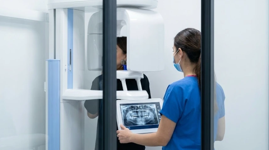
The question remains: is the radiation risk worth the diagnostic benefit? In our clinic in Turkey, we utilize the latest digital sensors, which reduce radiation exposure by up to 80% compared to old-school film.
For example, the radiation that comes from one dental X-ray is almost the same as the radiation that you get naturally from the sun during a three-hour airplane flight or by eating a few bananas (which have radioactive potassium-40). Hence, the potential risk to your health is very low, and the danger of a tumor left undetected or a bone infection becoming worse is something that changes your life completely.
FAQ: Direct Insights from Lema Dental Clinic
The reality is that pain is often the last symptom to appear. By the time a cavity hurts, it has likely reached the nerve. X-rays catch the fire when it’s just a spark, not a forest fire.
This is not a “one size fits all” answer. Dentist Polen Akkılıç and her team tailor this to your risk profile. If you have a history of frequent cavities, every 6-12 months is standard. If you have a “steel smile” with no history of decay, every 24 months may suffice.
Yes. While we generally postpone non-essential X-rays during the first trimester, the American Dental Association and the American Pregnancy Association agree that dental X-rays with proper shielding are safe. An untreated dental infection poses a far greater risk to the baby than a localized X-ray.
They are instrumental in finding tumors or cysts within the jawbone that a visual cancer screening would miss. It is one of our most powerful tools for early life-saving intervention.
Transillumination can help, but it cannot penetrate the high density of the jawbone or see “around the corner” of a deep molar root. There is currently no technology that replaces the clarity of ionizing radiation for bone-level diagnosis.
- American Dental Association (ADA). (2024). Radiographs/X-Rays: Clinical Recommendations and Safety Guidelines.
- Misch, C. E. (2017). Contemporary Implant Dentistry. Elsevier Health Sciences.
- Pauwels, R. (2020). Cone Beam CT of the Head and Neck. Springer Nature.
- White, S. C., & Pharoah, M. J. (2018). Oral Radiology: Principles and Interpretation. Elsevier.
- Yıldız, C. (2025). Digital Imaging Advancements in Modern Turkish Clinics. Journal of Istanbul Dental Sciences.