

Yes, decay can still occur
After a root canal procedure, leaving the clinic in Turkey releases a feeling of immense relief. Your agony has stopped, your infection has healed and your tooth is secured with a brand new crown. It’s almost as if the problem has been solved forever.
One question that we always get at our follow-up visits is: “Now that the nerve is gone, is this tooth indestructible?” The truth is a tough no.
Actually, a tooth with a root canal is probably more at risk of developing cavities than a living one. Not because the enamel is less strong but because the alarm system has been disabled. We, at Lema Dental Clinic, through our experience, observe that one of the main reasons why adult patients lose their teeth is due to what we call “recurrent decay” under their crown.
The “Helmet” Analogy
In order to grasp how a “dead” tooth can be affected by cavities, you need to consider what a dental crown is made of in an entirely different way.
Professor Doctor Coşkun Yıldız frequently repeats: Imagine a dental crown as a crash helmet. It is tough, long-lasting and made to protect against damage. However, the tooth underneath is still the “head”. If germs manage to get past the rim of the helmet (the margin where the crown meets the gum), the head inside can still be harmed.
The tooth under the layer of ceramic or Zirconia is still a biological material of organic nature. It still contains calcium and phosphate. If sugar and bacteria accumulate at the gumline, that organic matter will decay, no matter if it has a nerve or not.
Why It Is Called the “Silent Killer”
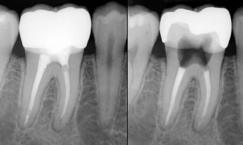
This is the nightmare situation. When a healthy tooth develops a cavity, it makes you aware of it. You get a sharp pain when water hits your mouth and a continuous ache after eating sweet stuff. The source of those sensations is worth it since it encourages you to visit your doctor.
In contrast, a root canal tooth is without a nerve. It has no sensation.
Here’s what usually happens: the patient decides to have a standard health check in Turkey, and feels great. However, even though Dentist Polen Akkılıç and her team find no external signs of the problem, an X-ray shows the tooth is practically gone because of a huge cavity. Until the patient notices that the crown is “loose” or has fallen out, decay has almost always reached such a level that the tooth can no longer be saved.
The culprit: Marginal Leakage
What drives bacterial invasion? This usually occurs at the “margin”—that tiny, almost invisible line where the crown meets your natural tooth.
In the course of many years, the dental cement can become loose. Besides, if oral hygiene is not kept at the right level, gum recession may lead to the exposure of the softer root surface underneath the crown. This results in a microscopic gap being formed. Bacteria are extremely small; therefore, they are able to get in through even a very small crack.
At Lema Dental Clinic, we minimize this risk by employing high-precision CAD/CAM technology to crown our work such that their fit is precise to the consistency of a micron. The more secure the fit, the less likely that bacteria can get in.
Comparing the Warning Signs
In the absence of pain as a reliable indicator, you have to find other indistinct signals. Below is a comparison of the manifestations of decay in a living tooth and a living tooth that has been treated.
| Feature | Living Tooth Decay | Root Canal Tooth Decay |
| Pain Level | Sharp sensitivity to cold/sweet | Zero pain (until infection hits bone) |
| Early Warning | Discomfort when chewing | Bad smell or taste in the mouth |
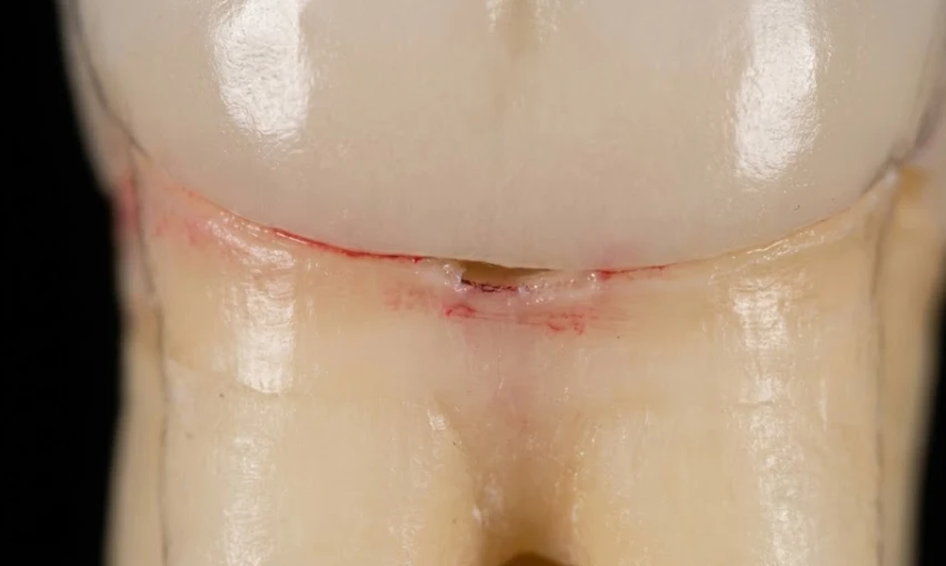
| Visual Signs | Dark spots on enamel | Red, inflamed gum at the crown’s edge |
| Structural Integrity | Usually remains stable early on | Crown may feel loose or snap off |
| Progression Speed | Moderate | Fast (often unnoticed until late stage) |
How to Protect Your Investment
You traveled to Turkey to save your smile, so protecting that investment is crucial. Since you cannot feel the decay, your defense strategy has to change.
- Floss the Margin: You must clean the area where the crown meets the gum daily. That is the entry point for bacteria.
- X-Rays are Non-Negotiable: Because these cavities are invisible to the naked eye, you need radiological evidence. We recommend a panoramic X-ray at least once a year.
- Watch the Gums: If the gum around a specific crown is always red or bleeds when you brush, it’s not just gingivitis. It could be a sign that the seal of the crown is failing.
FAQ: Answers from the Experts
It depends on how much “stump” is left. If we catch it early, we can remove the old crown, clean the decay, and make a new crown. But if the decay has eaten down to the root level, the tooth might need to be extracted and replaced with an implant.
Odor is a major red flag. If you floss around the crown and the floss smells terrible, it usually means bacteria are trapped under the crown, rotting the remaining tooth structure. You need to see a dentist immediately.
With perfect care, 10 to 15 years is a standard expectation. However, we see many last a lifetime. The longevity depends less on the porcelain and more on your hygiene habits.
Not always, but often. Sometimes the cement just fails. But frequently, when a crown pops off, it’s because the tooth structure underneath has softened from decay and can no longer hold the grip. Do not try to glue it back on yourself.
Material matters less than the “fit.” Whether it is gold, Zirconia, or porcelain, if the gap between the crown and tooth is wide, decay will start. We prefer Zirconia at Lema Dental Clinic because digital designing allows for an incredibly precise marginal fit.
- Aquilino, S. A., & Caplan, D. J. (2002). Relationship between crown placement and the survival of endodontically treated teeth. Journal of Prosthetic Dentistry, 87(3), 256-263.
- Tikit, T., & Attar, N. (2020). Longevity of crowns and fixed partial dentures: A scientific review. Journal of Dentistry and Oral Care, 6(1), 10-18.
- Goodacre, C. J., et al. (2003). Clinical complications with fixed prosthodontics. Journal of Prosthetic Dentistry, 90(1), 31-41.
- Ng, Y. L., et al. (2011). Tooth survival following non-surgical root canal treatment: a systematic review of the literature. International Endodontic Journal, 44(3), 175-189.
- Vire, D. E. (1991). Failure of endodontically treated teeth: classification and evaluation. Journal of Endodontics, 17(7), 338-342.