

3D scans ensure safe and precise implant placement.
Why a 3D Scan is Non-Negotiable for Your Dental Implants?
Imagine that you want to build a monumental modern skyscraper but you are only given a single instant photo of the land to base your project on. It is a fact that a camera can only lend the look of a surface to the picture, but the photographer is totally unaware of the underground piping, how deep the bedrock goes, or whether the soil can bear the weight of the building.
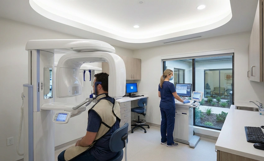
In restorative dentistry, it is almost like taking a health risk when you place implants without a 3D Cone Beam Computed Tomography (CBCT) scan. It would be a completely irresponsible and unprofessional act if you tried to place implants without 3D imaging in the entire dental field.
At Lema Dental Clinic, we don’t see smiles only as a set of teeth, but we also give them a thorough treatment as a complex architectural project. Unfortunately, the truth is, our jaws aren’t flat surfaces but a 3D world full of nerves, sinuses, and various densities of bones.
The 2D Limitation: Why “Standard” X-rays Aren’t Enough
For a long time, the panoramic X-ray was the most widely used. It is that flat, wide photo that captures your entire mouth in a single shot. Although the picture provides a general overview and gives some valuable information on your mouth, the most important limitation of it is that it misses the 3rd dimension or depth.
Imagine that one normal (2D) X-ray is just like a shadow puppet on a wall. You see only the outline but don’t know the thickness of the object or the distance from the surface. In preparation for complex implant surgery, Professor Doctor Coşkun Yıldız always requires, among other things, the exact millimeter of space available. At times, a 2D picture can be a “mask” by hiding bone deficiencies or bending the distance between vitally important structures.
Seeing the Unseen: Bone, Nerves, and Sinuses
The single most important phase of implant operation is below the gum line. The most significant part of the implant operation takes place underneath the gum line. This is what we observe at the office when we obtain a 3D scan:
First of all, bone volume and quality: we have to be certain of the “bedrock” to anchor the titanium post. A 3D scan indicates whether your bone is as hard as oak or as soft as balsa wood.
Secondly, nerve pathways: In the lower jaw, the inferior alveolar nerve functions as if it were a high-voltage cable. If the implantation procedure happens to touch it, the result can be permanent numbness. The presence of a 3D scan enables Dentist Polen Akkılıç and her team to mark off a “no-fly zone” around these nerves that is perfectly safe.
Last but not least, sinus cavities: In the upper jaw, your sinuses are right above the teeth. The sinus membrane is like a chicken’s eggshell. If the implant is too high without the guidance of a 3D scan, this delicate membrane may be torn.
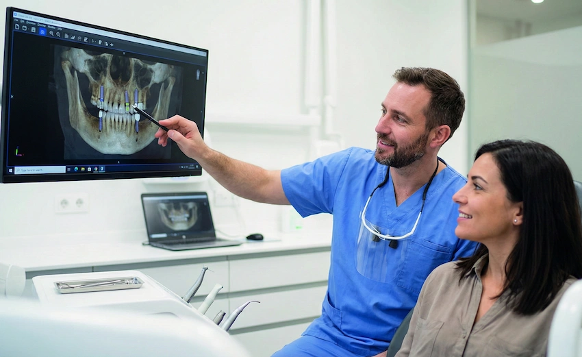
How We Use This Data at Lema Dental Clinic?
However, let us revisit the detailed actual operation. After the scan, the digital file is brought into the specialized physics planning software. It is the kind of software that makes the surgical team capable of “virtually” carrying out the operation long before the patient has ever sat down in the dentist’s chair.
Professor Doctor Coşkun Yıldız is known to say that the actual operation is the speedy implementation of a gearbox, the harnessing of i-pad, piloting, and launching from mission control “the lung surgery through the brain surgery screens”. The Istanbul facility is perfectly situated for such a process where one correctly prepared four-dimensional fractal model potentiates a high-resolution morphological version and production by minute and on stream, extensive, and additive integration of the mapping sanguine and other intermediates in cognitive functions. For our anatomy-specific purposes, we are already sure that the angle, depth, and size of the implant will be the best methods for the corresponding individual.
Comparing the View: 2D vs. 3D Imaging
| Feature | 2D Panoramic X-Ray | 3D CBCT Scan |
| Dimension | Two-dimensional (flat) | Three-dimensional (depth) |
| Bone Density | Estimated | Accurately measured |
| Nerve Mapping | Approximate | Precise to the millimeter |
| Surgical Accuracy | Moderate | Exceptionally high |
| Procedure Time | Longer due to intraoperative surprises | Shorter with pre-planned execution |
| Risk of Complications | Higher | Significantly minimized |
The Turkey Advantage: Technology Meets Expertise
Still, the question is why come all the way to Turkey for this? We are putting together the best technology worldwide with the clinical discernment of the experienced experts at Lema Dental Clinic. Having a scan is just one aspect; having a team like Dentist Polen Akkılıç and her team to analyze the data that comes from the scan is what fundamentally transforms the results.
We don’t only focus on “where the tooth should be.” We think about the implant as a means of supporting your facial contour, how it is going to work with your bite, and how to make sure it will still be there in 30 years, not just in 3 years.
FAQ: Your Questions Answered by the Experts
“Not in the slightest! The whole scanning process takes about 20 seconds and is absolutely painless and non-invasive. When it comes to radiation, modern CBCT scanners emit significantly less radiation than a medical CT scan – it’s more or less the same amount of radiation that the natural environment gives off in a few days.” – Lema Dental Team.
According to our clinical experience, yes. Even if you want only one tooth, the potential to bump a nerve or to find a lack of bone width still exists, and so we cannot afford to rely on guesswork. Above everything else, we want and insist on your safety.
We believe that precision is quite a natural human right and one of the most basic elements of the care standard we should all expect to receive. That’s why we have included thorough diagnostic tests in our treatment schedule so that your journey to Turkey can be a smooth one with no surprises.
Thanks to digital technology, we get everything so fast nowadays. It takes almost no time at all for the image to be processed, meaning that even during your initial consultation, both you and Professor Doctor Coşkun Yıldız can already discuss your digital ‘blueprint.’
Although it is acceptable to use it initially when you are having a remote consultation with us, once you are here, we will definitely need to do a fresh 3D scan. We require the most up-to-date, top-of-the-line data available so that your operation will be an absolute success.
- Bornstein, M. M., Scarfe, W. C., Vaughn, V. M., & Jacobs, R. (2014). Cone beam computed tomography in implant dentistry: A systematic review focusing on guidelines, indications, and protocol. International Journal of Oral & Maxillofacial Implants.
- Guerrero, M. E., Jacobs, R., Loubele, M., Schutyser, F., Suetens, P., & van Steenberghe, D. (2006). State-of-the-art on cone beam CT imaging for preoperative planning of implant placement. Clinical Oral Investigations.
- Harris, D., Horner, K., Gröndahl, K., Jacobs, R., Helmrot, E., Benic, G. I., … & Quirynen, M. (2012). E.A.O. guidelines for the use of diagnostic imaging in implant dentistry 2011. A consensus workshop organized by the European Association for Osseointegration. Clinical Oral Implants Research.
- Scarfe, W. C., & Farman, A. G. (2008). What is cone-beam CT and how does it work? Dental Clinics of North America.
- Tyndall, D. A., Price, J. B., Tetradis, S., Ganz, S. D., Kurmansky, C., & Brooks, S. L. (2012). Position statement of the American Academy of Oral and Maxillofacial Radiology on selection criteria for the use of cone-beam computed tomography in germ-line imaging and implant clinical dentistry. Oral Surgery, Oral Medicine, Oral Pathology and Oral Radiology.