

Buccal mucosal cancer is a cancer that arises from the lining of the cheek, and very often it can be seen as a non-healing ulcer, a thickened mucosal area, or an irregular lesion. The case is urgent for an early check-up since the cancer cells are capable of invading not only muscular tissue but lymph nodes as well. After a detailed examination of a patient at Lema Dental Clinic, Doctor Polen Akkılıç, with her staff,
feels is the first to detect abnormal tissue changes. Their well-organized routine permits each patient an immediate clinical contact, unambiguous support, and a securely followed-up plan resulting from the medical insight gained.
At the beginning, quite a few patients misinterpret their cheek lesions caused by biting, dental irritation, or local infections as something that just happened to them. Thus, those who eventually discover cancer in the deep structures of their bodies have lost the fight with the disease since their delay has made this terrible condition worsen. Prof. Dr. Coşkun Yıldız points out that the recognition of symptoms in stages is very important because the tumor that infiltrates and invades the deeper layers of the mucosa becomes more aggressive. Therefore advanced imaging, thorough palpation, and functional evaluation are employed by him to confirm the diagnosis, which precedes the spread of the disease in his clinical practice.
What Are the Characteristics of Buccal Mucosa Cancer?
It is basically a cancer of the cells lining the inner cheeks, which arise due to genetic alterations of a series of cells that have been irritated or exposed to substances that induce cancer.The mutations make the cells grow uncontrollably forming ones that are invasive and cancerous. Upon Polen Akkılıç‘s examination suspecting a malignancy, the dentist not only looks at superficial mucosal changes but also finds out whether deep structural parts are involved by checking if the lesion consists of squamous cell carcinoma, which is the most common subtype. Her team working together with her examines each change accurately to clinical staging and treatment planning which can be relied upon.
The issue turns out to be important in medical terms when the cancer cells break through the epithelial surface and spread to the submucosa or the muscles around it. Prof. Dr. Coşkun Yıldız explains that this pattern of penetrance is what directly determines the patient’s therapeutic options and chance for survival. His diagnosis method is all about locating this move from very local mucosal involvement to deeper tissue infiltration. Early differentiation between these ensures that each patient is receiving treatment that is needed, safe and effective.
Clinical Classifications of Buccal Mucosa Cancer
Doctors rely on the TNM system to classify cancer of the buccal mucosa. This system takes into consideration the size of the tumor, spread to the lymph nodes, and distant metastasis. Dentist Polen Akkılıç together with her team, notes the depth, width, and the invasion pattern of the tumor at the first visit to figure out the exact T stage. Their precise staging is the basis for every patient’s individualized high-tech treatment plan that is in harmony with global oncology standards. The patients receive solid information about their clinical stage and how it affects the treatment.
Besides that, the histopathological classification is a major determinant of the tumor’s aggressiveness. Professor Doctor Coşkun Yıldız works hand-in-hand with the pathology department specialists to examine the biopsy tissue samples. They make sure that the documentation of the tumor’s differentiation grade, keratinization pattern, and cellular atypia is done clearly. His well-organized investigation serves as an indicator of the treatment efficacy and overall prognosis.
Prevalence of Buccal Mucosa Cancer
The cancer of the buccal mucosa is mainly found in those areas where patients frequently use smokeless tobacco, drink alcohol, and have constant oral irritation. Dentist Polen Akkılıç meets with a large number of patients coming from such backgrounds and she instructs them on how their lifestyle choices affect the occurrence of cancer. She tells them how the long-term exposure of the mucosa to carcinogens gradually leads to DNA damage and eventually cancer.
The disease is more common in adults than in children. Professor Doctor Coşkun Yıldız thinks that the combination of risk factor exposure over the years and the aging-related changes of the mucosa makes it more susceptible. His practical knowledge points out that early detection in the high-risk group members is the key to longer survival and less harmful treatment results.
Presentation and Early Warning Signs of Buccal Mucosa Cancer
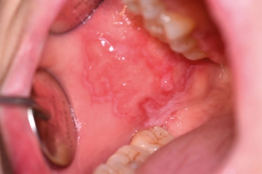
- Buccal mucosa cancer usually starts as an ulcer in the inner side of a cheek which doesn’t heal after two weeks. The lesion is generally a slightly elevated, uneven surface that extends its area gradually.
- A white patch (leukoplakia) or a red patch (erythroplakia) might be coming up in the region that is affected. These changes in the mucosa are the very first signs of dysplastic transformation and thus, they require recognition without delay.
- Localized cheek thickening is very often a hidden early sign and may even be slightly hard when you touch it with your fingers. The tissue may look swollen compared to the rest of the mucosa.
- The patients may feel a little pain while chewing or talking in the first stages of the tumor. When the tumor goes into the deep tissue, this pain becomes severe.
- A few patients might say that their mouth starts bleeding while brushing or chewing although no one provokes it. This bleeding indicates that the mucosa is very fragile and is made up of abnormal cells.
- If the cancer grows and starts pushing on the nerves, numbness or tingling in the cheek can be the result. This neurological symptom is an indication of the tumor’s deeper infiltration.
- The reduction of mouth opening (trismus) can be visible when the tumor involves the muscles around it. This sign is usually accompanied by an advanced stage of the disease.
What Causes Buccal Mucosa Cancer?
One of the main factors that led to the development of buccal mucosa cancer is the continuous exposure of the patient to tobacco, alcohol, viral infections, and mechanical irritation. Dentist Polen Akkılıç reports that she constantly comes across patients who have used smokeless tobacco or have experienced long-term mucosal trauma from the use of ill-fitting dentures. To her, it is very clear how the carcinogens from these sources break into the epithelial barrier and change the DNA of the cells, which leads to changes and eventually malignant tissue formation.
On top of that, alcohol consumption increases the risk factor by making the mucosa more permeable and lessening the tissue’s natural defense. Professor Doctor Coşkun Yıldız, in his turn, also speaks about the importance of the human papilloma virus, pointing especially to its high-risk strains that profoundly influence the behavior of the epithelial cells. His patient care reports involve advising people on ways to lessen the risk of the virus, giving up tobacco, and following protective oral health routines.
Major Risk Factors for Buccal Mucosa Cancer
- Long-term smoking of tobacco
- Use of alcohol
- Infection with HPV
- Bad oral hygiene
- Chronic cheek irritation
- Environmental carcinogen exposure
Diagnostic Protocols and Clinical Evaluation for Buccal Mucosa Cancer
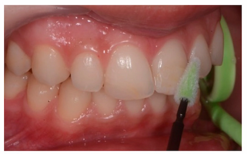
Diagnosis of cancer in the oral cavity requires a comprehensive method that involves examining the mouth, doing a biopsy, and taking pictures of the area. Dentist Polen Akkılıç follows a step-by-step method: looking at the lesion, feeling it with her fingers, measuring the lesion, and taking a photograph. After locating the area with suspicious tissue, they do a biopsy to check if it is cancer. The biopsy is very accurate in determining the grade of the tumor, keratinization, and the abnormality of the cells.
Professor Doctor Coşkun Yıldız does not stop at the local diagnostic examination and is using CT, MRI, or PET scanning to know the tumor size and whether the lymph nodes are affected. He conveys to the patients in an understandable way each radiological finding with which he is guided and how the result affects the treatment decision of the doctor. His working procedure is an insurance that every patient is very thoroughly staged and receives individualized treatment planning.
Diagnostic Tools for Buccal Mucosa Cancer
| Diagnostic Tool | Clinical Purpose | Value for Patient |
| Biopsy | Confirms malignancy | Ensures accurate diagnosis |
| CT Scan | Evaluates bone involvement | Guides surgical planning |
| MRI | Assesses soft tissue spread | Detects deep muscle invasion |
| PET Scan | Identifies metastasis | Predicts treatment direction |
| Oral Exam | Detects mucosal changes | Enables early diagnosis |
Treatment Modalities for Buccal Mucosa Cancer
The primary goal of the treatment is to remove the malignant tissue and stop the spread from happening any further. Dentist Polen Akkılıç and her team are involved with each patient in all the stages from pre-surgical preparation to postoperative care and oral rehabilitation planning. They help their patients by giving them detailed and clear oral hygiene, wound care, and functional recovery instructions so that the healing will be both effective and safe. Early cancer stages can be completely removed by surgery, thus cancer follow-up visits are the only necessary ones.
In the cases that are beyond the early stage, Professor Doctor Coşkun Yıldız comes up with combined treatment plans that include surgery, radiation therapy, and medication from the oncology field. To do so, he assesses tumor margins, lymph node status as well as patient overall health for not only safety but also therapeutic efficiency. His multidisciplinary approach decreases the chances of relapse and keeps the patient alive for a long time.
Risk-Reduction Strategies and Preventive Measures for Buccal Mucosa Cancer
Local risk reduction measures are to be taken only after the stopping of tobacco use; alcohol consumption should also be limited, and proper oral hygiene must be maintained. Dentist Polen Akkılıç through her organized counseling and preventive care stages, thus, patients become capable of protecting their mucosal tissue from continuous irritation and the intake of carcinogens. Besides, her preventive measures not only guarantee the oral health of the patient in the long run but also make them aware of the link between their daily habits and the danger of cancer.
Professor Doctor Coşkun Yıldız puts forward that visiting the dentist regularly and also the very early recognition of a non-healing lesion on the cheek should be taken very seriously. Apart from that, he also suggests general clinical practice vaccination against HPV, treatment of the chronic traumatic mucosa, and dietary supply as the most effective measures. Those who, particularly, adhere to these recommendations almost become free of the risk of cancer after a long period of time.
References:
- Chi, A. C., Day, T. A., & Neville, B. W. (2015). Oral cavity and oropharyngeal squamous cell carcinoma—an update. CA: A Cancer Journal for Clinicians, 65(5), 401–421. https://doi.org/10.3322/caac.21279
- Warnakulasuriya, S. (2018). Causes of oral cancer – an appraisal of controversies. British Dental Journal, 225(9), 841–848. https://doi.org/10.1038/sj.bdj.2018.906
- Johnson, N. W., Jayasekara, P., & Amarasinghe, A. A. H. K. (2011). Squamous cell carcinoma and precursor lesions of the oral cavity: epidemiology and aetiology. Oral Oncology, 47(4), 291–299. https://doi.org/10.1016/j.oraloncology.2011.01.009
- Shield, K. D., Ferlay, J., Jemal, A., Sankaranarayanan, R., Chaturvedi, A. K., Bray, F., & Soerjomataram, I. (2017). The global incidence of lip, oral cavity, and pharyngeal cancers by subsite in 2012. CA: A Cancer Journal for Clinicians, 67(1), 51–64. https://doi.org/10.3322/caac.21384
- Pindborg, J. J., & Reichart, P. A. (2018). Oral cancer and precancer. John Wiley & Sons.
Frequently Asked Questions About Buccal Mucosa Cancer
Buccal mucosa cancer develops in the lining of the inner cheek and needs to be diagnosed quickly by a professional healthcare provider. Treatment choices and survival rates can be positively impacted by a timely check-up.
It is very important that non-healing ulcers, persistent patches, or cheek thickening be looked at without delay. These signs can be those of cancer at an initial stage and therefore must be checked by a specialist.
Early-stage cancer is a good candidate for surgery and organized care, and it usually shows a positive response. For the advanced cases, the combined therapy is what helps to maintain life for a longer period.
One can say that tobacco, alcohol, HPV infection, and continuous irritation are the reasons that predispose to cancer development. So if you limit these risk factors the probability of cancerous changes will also decrease.
Biopsy and imaging are methods used by clinicians to confirm cancer and stage the disease. These instruments depict the entire tumor functioning.
Yes, in general, cancer could be local but extended local tumors may spread to lymph nodes or other organs. By early treatment, this risk is lessened and healing chances rise.
Lifestyle changes, preventive oral health care, and vaccination greatly lower the risk. Early examinations make it possible to recognize the lesions that cause concern in time.