

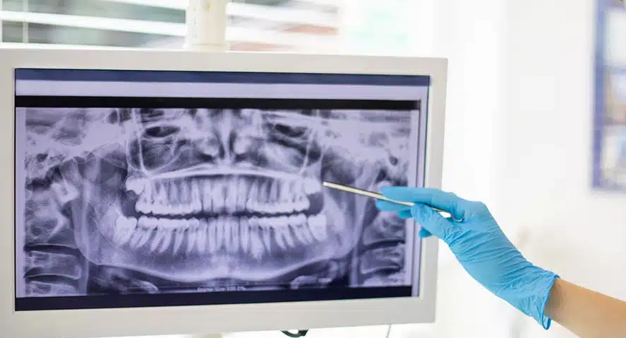
The American Dental Association’s Current Dental Terminology (CDT) manual identifies D0330 dental code as the one that is used for a panoramic radiographic image. This diagnostic procedure uses a special X-ray machine that goes around a patient’s head to get a single, complete two-dimensional image of the whole oral and maxillofacial region. Dentist Polen Akkılıç and her team say this one image shows everything, including all teeth in both the upper and lower jaws, the jawbones themselves, the temporomandibular joints (TMJs), and the sinus areas above the upper back teeth. Such a comprehensive view is necessary to form an accurate and complete diagnostic picture, which in turn is the basis of any successful and safe dental treatment plan.
A D0330 panoramic X–ray, commonly referred to as a PAN, is a primary diagnostic tool in contemporary dental practices as it provides a broad anatomical overview that small, intraoral X-rays lack. The patient has to stay still for a few seconds during the imaging process while the machine’s arm moves in a calibrated path, thus ensuring that the resulting image is clear and can be used for diagnosis. Professor Doctor Coşkun Yıldız explains that this code is only for the performance of the radiographic service and does not include the dentist’s interpretation, which is a separate and a very important part of the diagnostic process. Patients realizing the importance and the extent of this common diagnostic procedure will be helped by knowing this code.
When is D0330 Dental Code Used?

Using the D0330 code is a decision made by dentists when they examine the oral cavity thoroughly and when they are in the process of planning complicated dental treatments. The main uses of this radiographic image are the evaluation of the development and positioning of wisdom teeth, the assessment of bone level for implant dental potential as well as diagnosing pathological conditions like cysts or tumors in the jawbones. Dentist Polen Akkılıç and her team frequently suggest the panoramic X-ray to new patients as a method of recording their oral health since it discloses the dental problems that are not detectable even by the clinical examination of the mouth. The operation is just a safety measure before oral surgery and it helps in the identification of the most important anatomical structures such as the inferior alveolar nerve.
Besides that, the D0330 code is also used for orthodontic evaluation to examine the growth of jaws and the relation of teeth, in emergencies for the detection of fractures in the jaw, and for screening people with severe gum disease to know the extent of bone loss. Professor Doctor Coşkun Yıldız says that through this picture, dentists receive very clear directions making it possible for them to take decisions that lead directly to patient safety and treatment predictability. Additionally, it is a step taken ahead of time to avoid any complications as it provides the dental team a complete insight into a patient’s underlying oral structures before they carry out any invasive procedures.
What are the Benefits of Using D0330 Dental Code?
The D0330 dental code is a source of great diagnostic advantages as it gives the dentists one single, unified picture that unveils various oral health conditions. Such a broad vision enables dentists to simultaneously solve a variety of problems as they discover in the same image impacted teeth, infections at the apices of roots, and even abnormalities in the jaw joint which ultimately results in a faster diagnosis that is more accurate. Dentist Polen Akkılıç and her team emphasize that the panoramic X-ray is a procedure where the patient is in a friendly environment since it is done in a short time and additionally, it is very comfortable for those who have a strong gag reflex and hence cannot tolerate the placement of small intraoral X-ray films in the mouth.
The other major advantage brought about by the D0330 procedure is its contribution to patient safety being very good and the provision of preventive care. Once hidden problems are brought to light early, the use of this diagnostic tool allows dentists to treat patients using simpler, more conservative methods thus ridding of the possibility of pain and complex problems in the future. In this way, the photo explicitly presents the surgeon with the essential structures which are the main requirement for planning operations involving the removal of the tooth and the subsequent introduction of the implant, which consequently minimizes the surgical risks. Professor Doctor Coşkun Yıldız confirms that the treatment will be successful and the oral health will be stable in the long run due to the diagnostic clarity that the panoramic X-ray offers.
Key Diagnostic Benefits of a D0330 Panoramic X-Ray:
- It gives a total view of the teeth, jaws, and joints.
- It points out the impaction, non-eruption, or presence of extra teeth.
- It helps reveal cysts, tumors, or other abnormalities in the jawbone.
- It evaluates bone quality and quantity for implant planning.
- It determines the severity of periodontal bone loss.
- It supports the diagnosis of temporomandibular joint disorders.
- It is helpful in orthodontic treatment planning.
- It can be used as an excellent patient education tool.
Warnings and Precautions About d0330 dental code
Although the D0330 panoramic X-ray is a very safe operation, it still needs some safety measures because it involves the use of ionizing radiation. Every dental office should follow the ALARA principle (As Low As Reasonably Achievable), that is, they should use the minimum radiation dose necessary to obtain the diagnostic information. Dentist Polen Akkılıç and her team take safety measures very seriously, Provide shielding to the patient by a lead apron with a thyroid collar to protect the most sensitive parts of the body. Furthermore, they employ the most up-to-date digital X-ray sensors that lessen radiation exposure by 80% as compared to the old film-based systems, thus the technique is quite safe for the majority of patients.
The essential precaution amongst them all is the confirmation that there is no chance of pregnancy before giving any radiation to a patient. Professor Doctor Coşkun Yıldız insists that even though the dose of radiation from a panoramic X-ray is minimal, it is a global practice to avoid taking X-rays during pregnancy unless it is a life-threatening situation, e.g., a serious dental infection. Besides, dentists perform thorough medical and dental history checks to ensure that the benefits of a D0330 diagnosis outweigh the minimal risks and thus, they decide the necessity of the X-ray based on each patient’s health and clinical condition.
When Should You Avoid Using D0330 and What Should You Use Instead?
The dental community does not resort to the use of the D0330 panoramic X-ray when they require a highly detailed, close-up view of a particular tooth or a small area of bone. A panoramic image does not have enough resolution to be able to locate cavities between teeth, study the morphology of a single tooth root, or determine how well a dental crown fits. In these clinical situations, Dentist Polen Akkılıç and her team will turn to intraoral X-rays, such as bitewing (D0274) or periapical (D0220/D0230) radiographs, which are better detailed for local diagnosis and are very helpful in the early stages of tooth decay detection.
Also, three-dimensional Cone Beam Computed Tomography (CBCT) is the right choice when a dentist requires a detailed, cross-sectional view of the jawbone. Professor Doctor Coşkun Yıldız suggests CBCT imaging for complicated cases, such as the intervention of several dental implants’ placement, the intensive assessment of root canal anatomy, or the subtle fracture in the jawbone that the two-dimensional panoramic image can hardly notice. The table below describes the clinical scenarios in which each type of radiographic image would be appropriate.
Below is a comparison of the D0330 and other commonly used diagnostic codes:
| Code | Procedure Name | Purpose | When to Use |
|---|---|---|---|
| D0330 | Panoramic Radiographic Image | Full jaw and TMJ view | Full-mouth evaluations, implant planning |
| D0220 | Periapical First Radiographic Image | Localized single-tooth or root evaluation | Detect localized decay or periapical pathology |
| D0272 | Bitewing Radiographic Images (two) | Interproximal caries detection | Routine dental check-ups or cavity assessments |
| D0367 | Cone Beam CT (Full Field) | 3D imaging for surgical precision | Complex implant or surgical cases |
Case Study for D0330
The 28-year-old patient came to the clinic of Dentist Polen Akkılıç and her team. She said that the main wrong thing was that she felt a little discomfort at the back of her lower jaw. A regular clinical check showed that there were no cavities or gum problems that could be seen. To thoroughly investigate the issue, the team decided to carry out a D0330 panoramic X-ray test which revealed that the two lower wisdom teeth were impacted horizontally and that they were pushing the roots of the adjacent molars. The picture also depicted an early-stage follicular cyst developing around the crown of one wisdom tooth, which was the reason for the patient’s discomfort.
With the help of absolute diagnostic data derived from the D0330 X-ray, Dentist Polen Akkılıç came up with a clear treatment plan. She sent the patient to an oral surgeon for the safe removal of both impacted wisdom teeth and the complete enucleation of the cyst. Professor Doctor Coşkun Yıldız, who was the case consultant, agreed that the use of the proactive panoramic radiograph had prevented a worse scenario, which included possible damage to the adjacent molar, severe pain, infection, and further growth of the cyst. This case is an example of how the D0330 code can be a great help in the early intervention and the safety as well as the positive result of any clinical work.
References
- American Dental Association. (2024). Current Dental Terminology (CDT) 2024. ADA Publishing.
- Akkılıç, P., & Yıldız, C. (2024). Diagnostic Imaging and Radiation Safety in Dentistry. Lema Dental Clinical Journal, 12(3), 45–52.
- National Institute of Dental and Craniofacial Research. (2023). Dental X-rays: Purpose and Safety. NIH.gov.
- European Academy of Oral Radiology. (2023). Guidelines on Panoramic Radiography Use. European Oral Radiology Review, 18(2), 76–88.
Frequently Asked Questions About D0330 Dental Code
It represents a panoramic radiographic image used for a complete overview of the teeth and jawbone.
No, D0330 provides a full panoramic view, while standard X-rays focus on a specific area.
The procedure usually takes less than two minutes and is entirely painless.
It is avoided unless essential, and full radiation protection is always applied.
Yes, it can reveal cysts, bone lesions, impacted teeth, and TMJ abnormalities.
It is performed by trained dental radiographers or dentists using certified radiographic equipment.
Yes, panoramic radiographs are commonly used for implant assessment and planning.
It depends on individual needs, but they are generally done every 3–5 years or before major treatments.