

Sometimes understanding dental health can be a little confusing, especially when it comes to the machines your dentist uses. In fact, dental X-rays are one of the main tools in modern preventive dentistry. They are necessary because they offer a look into the parts of your mouth that cannot be seen, even with a visual exam. This ultimate guide will give you the power of understanding with clear, precise, and comforting pieces of information.
We will look at the different dental X-ray types, explain the factors that determine their frequency, and the safety profile at a glance. Our objective is to convert your knowledge from confusion to assurance so that you understand and trust the very care that keeps your smile healthy for years to come.
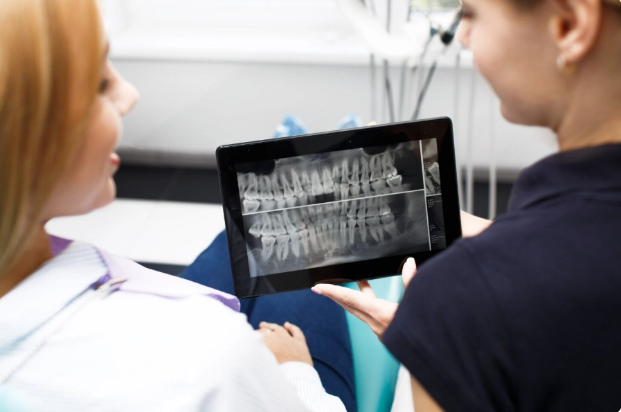
How Often Do You Need Full Mouth Dental X-Rays?

A full mouth series of dental X-rays, or FMX, is the most in-depth radiographic evaluation in dentistry. The pictures in this collection depict the entire oral cavity in a detailed way. Each tooth, from crown to root, the bone that supports the teeth, and even the tissues around it, is all shown here. The full mouth series is what the dentists suggest as an absolute necessity for new adult patients. This is how the initial oral health status becomes recorded in the patient’s file.
Diagnostic clarity is the first and most important thing that stems from this investment: the existing conditions, for example, cavities going on under the gums, bone loss, abscesses, cysts, or developmental anomalies can be easily figured out in this way because they are detected normally.
Your dentist will advise you to have a new full mouth series every three to five years. This time span is chosen according to the typical rates of progression of most common dental conditions, thus dentists can plan ahead for timely treatment. The huge diagnostic power of an FMX is based on it showing differences over time, therefore giving a comparative baseline which cannot be seen in bitewing X-rays. For example, it helps the dentist to be very accurate in following up on the slight changes in the bone density and levels that are caused by periodontal disease, checking the condition of roots, and looking for any pathological changes in the jawbone.
What about the question: How often do you need dental X-rays? The decision concerning how often one should take minimally invasive dental radiographs is the result of a personalized clinical judgment that is very carefully adjusted to an individual’s oral health status, age, and particular risk factors. The dentist follows a well-known international standard called ALARA (As Low As Reasonably Achievable) principle, meaning that the least possible amount of radiation is used for a medical purpose and that all X-rays taken are medically justified. In the case of a healthy adult who is a low caries risk, practicing good oral hygiene, and without periodontal disease, the biting-wing interproximal X-rays can be taken every 18-24 months. These are the types of radiographs that are aimed at the shortest distance between two adjacent teeth, in this way, dentists can detect the beginnings of decay in a place that is unseen by the naked eye.
Yet, if the risk factors assessment for you indicates a higher vulnerability to dental diseases, then your dentist will decide and suggest more frequent visits for monitoring purposes. A person who has been suffering from the recurrence of caries, diagnosed with periodontal disease, his/her diet packed with sugar, and has a dry mouth problem (that is usually the side effect of some medications) as well as a complex history of dental work, will benefit more from bitewing X-rays once every six to twelve months. Such an initiative is far from being over the top; on the contrary, it is a standard approach to higher-risk situations and thus a way of managing them. It lets the dental practitioner find the cause of the problem when it is minimal and solvable, most probably by using a simple filling rather than performing a root canal or a crown.
Such a method keeps your natural teeth intact, alleviates pain, and is the most cost-efficient way of oral health; thus, X-rays are a great investment for your well-being.
How Often Do You Need Panoramic Dental X-Rays?
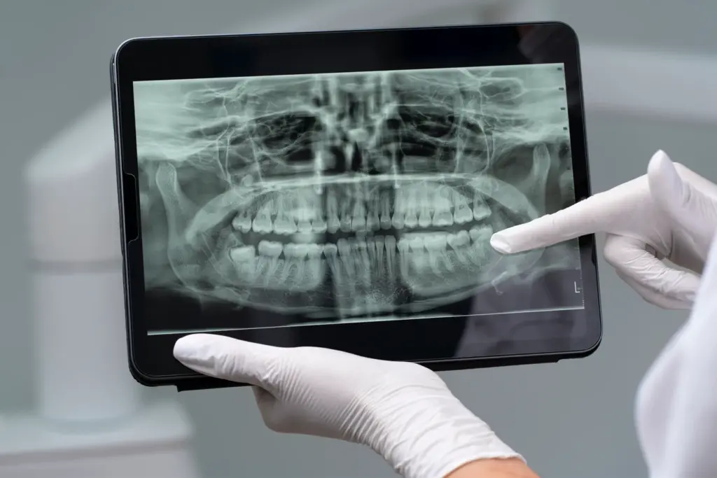
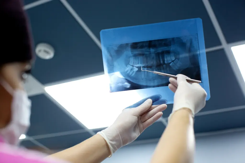
A broader panoramic dental X-ray, or Panorex, is an important and one-of-a-kind image that offers a wide, single view of your oral and maxillofacial structures. Intraoral X-rays are narrow and focused on a few teeth only, whereas the panoramic image is like a vista that shows all the upper and lower teeth, both the jaws, temporomandibular joints (TMJs), and the maxillary sinuses. Dentists and specialists depend on this large picture for a thorough examination and the planning of complicated procedures. In brief, the major uses include checking out the development, location, and removal if necessary, of the wisdom teeth; creating a thorough plan for orthodontic treatment like braces or clear aligners; figuring out the quality and quantity of the bone for dental implant purposes; and, determining the cause of the jaw pain or facial injury.
In fact, the frequency of panoramic X-rays is something that is very far from being on a routine level because it is usually done for a specific diagnostic purpose only. Generally speaking, a panoramic X-ray can be considered as a general checkup tool for saving the overall health and development of the jaw and therefore it is recommended every five to seven years. Nevertheless, your dentist will surely suggest a new panoramic picture every time you embark on a new and major dental treatment phase. For example, if you are going to be an orthodontic patient one day, either as a teen or adult, the current panoramic X-ray would be a must-have for your orthodontist in order to safely plan your tooth movement. Similarly, if you want to replace the missing teeth with dental implants, the first test he will do is a panoramic image to get a rough idea of your jawbone.
The idea behind targeted usage is that it assures every panoramic X-ray keeps giving the utmost clinical utility, thus supplying all the comprehensive data your dental team needs for the accurate, safe, and successful completion of complex treatments.
Dental X-Rays: How Often Is Safe?
Safety is a very important issue, and modern dental radiography can be counted as one of the safest and most reliable diagnostic practices due to major technological improvements and strict safety instructions. One of the greatest changes that come to a large extent due to the use of digital X-ray systems is the very significant decrease in patient radiation exposure (up to 90% compared to traditional film-based X-rays). The security of you, the patient, is also supported by the necessary protective steps, e.g., a leaded apron that is wrapped around your torso and a thyroid collar that goes over your neck and effectively protects your body from any scatter radiation.
To give a good idea of the extremely low-level radiation that is involved, think about that the set of four routine digital bitewing X-rays will give you a dose of approximately 0.005 millisieverts (mSv). If we put that amount of radiation into context, the regular person living in the US is exposed to roughly 3.1 mSv of background radiation annually, which comes from natural sources in the environment, such as radon gas and cosmic rays from space. This means that the exposure you’ll get from a dental X-ray is less than the plasma radiation exposure you would get if you were to stay inside your house for 24 hours. The ALARA principle is what your dentist is committed to, and thus, every X-ray is taken solely for a specific and beneficial reason. If dental X-radiographs are done according to your personal health circumstances, then these examinations pose an extremely low risk and provide a very essential diagnostic benefit.
How Often Do Adults Need Dental X-Rays?
The timetable of adult dental X-rays is dynamically and cautiously adjusted to be a part of your ongoing care and to be in line with your ever-changing oral health needs. For a healthy young or middle-aged adult with a low risk profile (good oral hygiene practice, a diet low in sweet foods and drinks, and no active diseases), the regularity of routine bitewing X-rays is set to be once every 18 to 24 months. This time interval is very suitable to controlling the areas tightly directed between the posteror teeth for early decay signs and for checking the artificially-restored teeth, such as fillings and crowns, thus verifying that they remain sealed and recurrence of decay-free.
Older adults or those with risk factors are the ones whose need for regular X-ray check-ups is likely to be increased as a result of wise decision-making. Such adults as moderate to high risk of dental diseases and individuals with frequent caries, diagnosed periodontal disease, and conditions like dry mouth (xerostomia)—a common side effect of hundreds of medications—will greatly benefit from the rehabilitation of their dentition through frequent visits to a dental practitioner. For these people, the role of radiographs every six to 12 months is a standard preventive and indispensable measure. More frequent checks give a chance to dentists to cover the higher risk of root caries (thus, gingival recession and subsequent root exposure), decay recurrence close to the old dental works, and the acceleration of periodontal disease. Fierce and customized monitoring is the leading sign of good dental care, which is at the same time, a brilliant way of detection and intervention planning, conservative in nature, and focused on preserving patients’ natural dentition for a lifetime of function and comfort.
Pediatric Dental X-Rays: How Often?
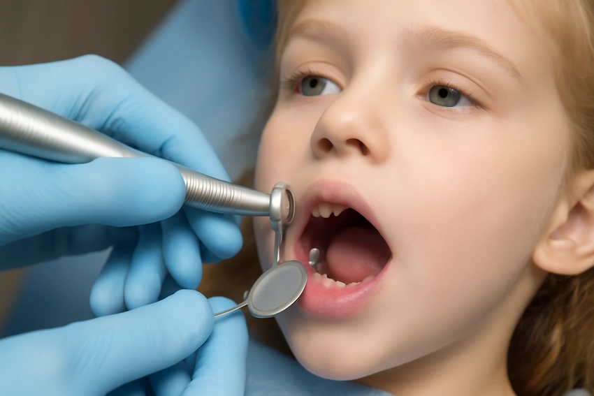
Kids’ dental X-rays are a necessary and invaluable weapon in the fight for the health and the correct development of your little one’s smile. A visual check is far from enough to look into the most important oral health aspects of a child and to figure out the position as well as the development of the unerupted permanent teeth lying under the gums; the presence of decay forming in the tight contacts of the baby teeth; or the first signs of infection or developmental problems. The American Academy of Pediatric Dentistry (AAPD) gives a clear set of rules and suggests that the frequency of X-rays be dictated by the child’s individual caries risk assessment. If a child is full of cavities, he/she might need a radiographic snapshot twice a year to be checked for new cavities and early treatment. A child with low risk might only take X-rays every 12 to 24 months which is just a routine screening.
Most likely, the first X-ray is a recommendation when the primary molars in the back of the mouth have grown in and are touching each other, usually between the ages of four and five. The first imaging is very important for setting a baseline and checking the interdental decay. Children’s dental clinics are especially fitted out with the fastest digital X-ray sensors and lead aprons and thyroid collars sized for children, ensuring the absolute highest safety level. Picking up the hidden cavity early—thus enabling a small, simple filling instead of the painful side of pulpotomy (baby root canal) or extraction—greatly outweighs the negligible risk. These snaps are adamant for the safe eruption of the permanent teeth and the safety of your kids against dental pain, thus they are a proactive and indispensable part of their wellness.
How Often Should a Teen Get Dental X-Rays?
Adolescent dental years are a time of major changes in dentition and thus, teens’ preventive care must include regular radiographic checks. At such a time, the last permanent teeth, excluding wisdom teeth, are coming out, and the jawbone is growing its last. Dentists typically recommend a 12-18-month interval for teenage X-rays to be able to keep an eye on this dynamic environment. Bitewing X-rays are still very important in spotting decay in the newly erupted permanent molars, which are extremely vulnerable to decay because of their deep grooves and fissures, and also mainly because of the changes in dietary and oral hygiene habits that usually occur during the busy years of life.
Besides, the teen years are mainly the time when orthodontic evaluation and wisdom tooth assessment happen. In many cases, a panoramic X-ray is suggested during this time to give one, overall, clear image that discloses the presence, location, and growth of all four wisdom teeth, the roots of all permanent teeth, and the jaw’s overall structure. Such information is beyond measure in the process of determining the need for braces, planning extractions, and foreseeing potential problems before they manifest in the form of pain or crowding. Like other patients, dental professionals utilize the most modern digital X-ray technology and strong protective measures to make the process very safe for teenagers. Regular and planned X-ray use liberates your dental team to get your teenagers through their critical developmental period, thus guaranteeing they will have a healthy, straight, properly functioning, confident smile that is ready for adulthood.
Sources
- American Dental Association (ADA). (2023). Dental Radiographic Examinations: Recommendations for Patient Selection and Limiting Radiation Exposure. U.S. Food and Drug Administration.
- Health Physics Society. (2021). “ALARA – As Low As Reasonably Achievable.”
- U.S. Food and Drug Administration (FDA). (2023). “Medical X-Rays: Guidance for Patients and Caregivers.”
- American Dental Association (ADA) Council on Scientific Affairs. (2023). “The Use of Dental Radiographs: Update and Recommendations.”
- American Academy of Pediatric Dentistry (AAPD). (2023). “Prescribing Dental Radiographs for Infants, Children, Adolescents, and Individuals with Special Health Care Needs.” The Reference Manual of Pediatric Dentistry, 334-339.