

The Role of Dental X-Rays in Oral Cancer Diagnosis: How Can They Detect Early Signs?
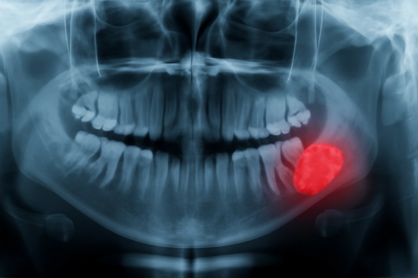
Dental X-rays are the primary means through which oral cancer is prevented in its very early stage by locating very subtle changes. These radiation images give a close-up of the teeth, gums, and bone involved around the teeth and the oral health specialists are able to find these abnormalities in the very earliest stage by detecting tumors, cysts, and unexplained growths. For the group of patients liable to oral cancer, dental X-rays might turn out to be a very efficient preventive measure, leading the way towards identification of phenomena such as alterations in bone density or the presence of abnormal tissue. The notion of early findings is so important because at that moment there is an opportunity for very prompt intervention and the chances of treatment being successful are significantly increased.
Moreover, dental X-rays also prepare the ground for the future by setting a standard for comparison and thus by offering the possibility of detection of gradual changes that occurred since the last visit apart from revealing the immediate visual signs of disease. In case of abnormality, the dentist will be able to watch the development of the issue through frequent check-ups, making sure that the problem is recognized in the initial stages. This applies especially to people who have a higher risk such as those who have been smoking or consuming alcohol excessively over a long period of time. The earliest finding of oral cancer by dental X-rays can bring about very fast and consequently more effective treatment, thus, very often, patients may have better outcomes.
Dental X-Rays in Early Oral Cancer Diagnosis: How Critical Are They?
Dental radiographs are indispensable instruments in the early detection of oral malignancy and provide the first indication of various abnormalities. They are the absolute necessities for the following reasons:
- Firstly, dental X-rays uncover the changes which indicate cancer at the initial stages to oral cancer, these may be abnormal growths, bone structure alterations, or lesions. Detecting these changes at the stage, dentists can direct their patients to undergo a series of tests and get treatment in time.
- Secondly, dental X-rays present a non-invasive and accessible screening method for oral cancer, thus are available for most patients. In contrast to more invasive procedures, dental X-rays are very much time-saving, easy-to-do, and may become a part of routine dental visits.
- Thirdly, X-ray images, especially panoramic X-rays, cover a relatively large area of the whole mouth, teeth, and jawbone, thus depicting the entire picture. This detailed image helps the doctor find places where cancer may be hiding, and there is no doubt that these areas have not been visible under the visual examination pattern.
- Fourthly, X-rays are a necessary first step in identifying suspicious regions, though they cannot alone be used to confirm oral malignancy. They are used by dentists to pinpoint areas that need biopsy and to decide the extent of tissue removal, thus the presence of cancer is confirmed properly and gets thorough investigations.
- Fifthly, intervention at an early stage and a positive outlook is the direct outcome of oral cancer detection through dental X-rays. This is achieved through less invasive methods of drug administration and the patient’s general well-being improvement, giving a brighter prospect for the almost complete recovery situation.
- Last but not least, Dental X-rays-Musical acts (the duo) comprising of the oral malignancy and diagnostic assistants, are supplemented with one another’s capabilities and resources. Combining this with a patient history’s taking and a visual examination, it provides the most comprehensive pathway in terms of cancer detection, its reliability is improved.
Promoting dental X-rays to be a method of routine screening, oral cancer detection would be early and thus facilitated to a large extent and those who are at risk would be under the close and careful monitoring by the healthcare professionals, leading to the better outcomes.
Oral Cancer and Dental X-Rays: What Types of X-Rays Are Used for Cancer Screening?
Any dental X-ray can be utilized to find mouth cancer, but each of them has different advantages depending on which part is to be checked. For example, panoramic X-rays not only show the teeth but also the gums and the bones of the jaw, thus, providing a complete view of the mouth.This kind of total image is able to make large tumors and other abnormal growths that are matted deep and, therefore, may not be visible even fished out. In addition to that, panoramic X-rays allow dentists to measure the entire oral cavity and then it becomes very easy to locate the abnormalities of the teeth as well as the tissues that are close to them.Due to its feature of gathering various kinds of data in a single shot, this X-ray is usually the one that is first used in an oral cancer examination.
Bitewing X-rays in combination with periapical X-rays are not the most efficient ways to locate oral cancer, but they can be very helpful in this area. The main function of Bitewing X-rays is to find those areas where the teeth get the most wear, thus, they are the most capable of detecting dental caries. If cancers through observation of bone or tooth structure changes were present, then this would be an aid in the indirect indication of oral cancer.Periapical X-rays focus on one single tooth and the area around it and the structure, thus giving more details of the root and the bone. Such a highly targeted method can detect changes in the bone structure that may have been caused by cancer at a very early stage, thus a more focused approach in identifying oral health issues.So, by using these different types of X-rays, no part of the oral cavity is left without a check.
Oral Cancer and Dental X-Rays: New Technologies for Early Diagnosis

The improvements of dental X-ray techniques have impacted positively on the precision and trustiness of oral cancer detection, which are primary goals to achieve in recent times-hence the successful example of digital X-ray. They provide photograph quality at least 3 times better than the already existing film camera. The finding of oral cancer in its incipient stage is feasible through digital X-rays due to the clarity of the structures shown by it. In addition, the digital mode of operation is less harmful in terms of radiation dose, thus making the digital X-rays an excellent alternative, especially for patients requiring frequent screenings. Additionally, the fastness of digital X-ray operate provides a prompt image review by doctors hence the whole diagnostic procedure is sped up and the patients get timely treatments.
Cone-beam computed tomography (CBCT) is another advanced technology that is changing the paradigm of oral cancer diagnosis. CBCT is a 3D imaging method that offers remarkable detail and precision and reveals much more than regular 2D X-rays can show. With CBCT, dentists have a keen look at the bones, teeth, and soft tissues with an added layer of detail, and thus can discover different suspicious areas that are potential sites for cancer at an early stage. This technique helps in the detection of hidden tumors or abnormal areas that are virtually impossible to find with regular X-ray. When the oral cavity can be seen in 3D, it not only helps early detection but also makes treatment planning efficient hence CBCT is a valuable weapon to fight oral cancer.
Oral Cancer Screening with Dental X-Rays: When Should They Be Performed?
An oral cancer screening with dental X-rays should be performed at a time that is closely related to a person’s indvidual risk factors. People who have a family history of oral cancer or are smokers should start their regular screenings as early as possible. Generally, one can say that such cancer screenings should be carried out with X-rays as part of a routine dental check-up that normally takes place once every one to two years for the average adult. Nevertheless, those individuals who have a higher risk may be required to undergo the test more frequently. Depending on what the dentist advises and the medical history of the person, these patients may have to take the X-ray examination even more than once in a year.
Oral cancer treatment starts with its timely discovery, and one of the measures that can help is a dental X-ray. The mentioned preventive measure cannot be missed by high-risk subjects because the first intervention thereupon can raise greatly the chances of recovery. Dentists should evaluate patients’ risk factors and decide the intervals for their checks so as to keep very close surveillance on at-risk individuals for any symptoms of oral cancer. Besides, the inclusion of dental X-rays in regular check-up visits can be the patients’ way of prevention.
Early Detection of Oral Cancer: How Reliable Are Dental X-Rays?
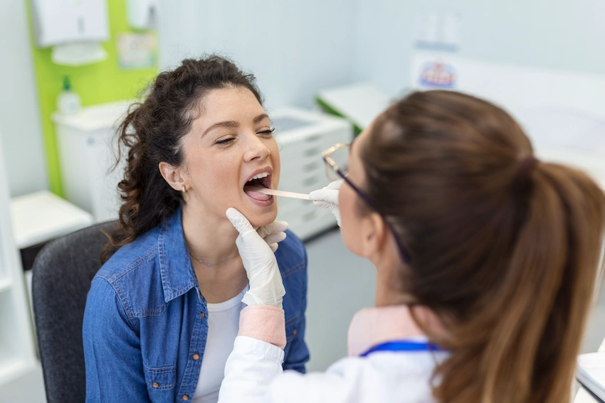
Dental X-rays reveal a lot about early cancer symptoms in the mouth, especially when they are within a patient’s full medical examination strategy. On the X-rays, tumors, cysts, and cancerous changes in the bone can be seen, which indicate oral cancer. That is why doctors take X-rays only as an initial step towards understanding a problem.
The efficiency of dental X-rays depends on the clarity of the pictures and the dentist’s skills in recognizing the problems. The use of modern digital and 3D technology for dental X-rays has increased the precision of such X-rays and has almost eliminated doubts associated with the early-stage cancer issue. Despite that, a patient should be physically examined and interviewed, along with X-rays taken, to get a complete overview of his/her case. If used regularly, dental X-rays can be a strong weapon against cancer in its early stages, but their function can only be successful when they are accompanied by other diagnostic tools.
Dental X-Rays and Oral Cancer: Raising Awareness and Preventing Steps
The very first point is to make the information of dental X-rays as an oral cancer-detecing tool known to the public to facilitate early detection rates of cancer as well as fast health diagnosis. The summarized points are as follows:
- Education on Oral Cancer Risks: Firstly the campaigns should help individuals understand different factors that can cause oral cancer eg smoking eating factors, utilization of heavy drinkers and lineage of the disease. In this case people will be aware of saving their oral health through safe lifestyle choices and get committed to the importance of dental visits.
- Promote Routine Screenings: Create a demand for regular dental check-ups where people would undergo X-ray screenings. A routine dental exam encompassing an X-ray is a must in any early-phase oral cancer detection without an in-sensibles situation.
- Targeted Screening for High-Risk Groups: The message of continuous detailed examination should be loudly told to those among the risk of oral cancer like habituated smokers, drinkers, and lineage of the cancer-affected families. Nowadays a lot of these people are acquainted with X-ray has more cancer symptoms than others may have.
- Collaboration Between Dentists and Other Healthcare Providers: A collaboration between dentists and doctors could continuously help people at-risk write their care and look at cancer from a broader perspective. One cannot oppose that doctors can imply to patients with such disorders the need to go to dental experts for oral cancer checkups during general health monitoring.
- Lifestyle Modifications: Helping the public through education on the changing of lifestyles that would help stop oral cancer via practices like smoking cessation, alcohol use restriction, great oral hygiene. Supporting these interventions can go a way to bring oral healthcare needs to the fore.
- Increase Awareness Through Social Media and Community Outreach: Utilization of social media-scale and community engagement activities will act as a pacemaking tool for quicker and easier diffusion on oral cancer-related information, in which they do great in terms of oral health care. Influential oral health promotion materials like stories and stats could be made accessible to users to urge their taking responsibility for their oral health.
Types of Dental X-Rays Used for Oral Cancer Detection
| Type of X-ray | Purpose | Benefits |
| Panoramic X-ray | Provides a full view of the mouth, teeth, jaw, and surrounding areas. | Detects large tumors or abnormalities not visible during a regular exam. |
| Bitewing X-ray | Shows detailed images of the upper and lower teeth. | Helps detect cavities and other minor issues, potentially indicating early cancer signs. |
| Periapical X-ray | Focuses on individual teeth and surrounding bone. | Identifies changes in bone structure or early signs of abnormal growths. |
| Cone-beam CT (CBCT) | Provides 3D imaging of the oral cavity. | Offers precise, three-dimensional views of bone structures and soft tissues. |
References:
- Arboleda, A., & Saitta, J. M. (2020). The role of dental imaging in early detection of oral cancer: An overview. Journal of Clinical Dentistry, 31(2), 89-96. //doi.org/10.1002/jcd.10234
- Ghaffari, M., & Shahravan, A. (2019). Dental radiology in the early diagnosis of oral cancer: An assessment of X-ray techniques. Oral Health & Preventive Dentistry, 17(3), 217-223. //doi.org/10.3290/j.ohpd.v17i3.80009
- Nandini, R., & Chawla, S. (2021). Advances in dental X-ray technology for oral cancer detection. Dental Research Journal, 18(4), 210-217. //doi.org/10.4103/drj.drv18i4.3217
- Tandon, S., & Singh, R. (2020). Panoramic radiography in oral cancer detection: A clinical approach. International Journal of Oral Science, 12(1), 22-29. //doi.org/10.1038/s41368-020-0031-4
- Wong, T. Y., & Lee, A. H. (2021). Early detection of oral cancer using dental X-rays and adjunctive technologies. Journal of Cancer Detection & Prevention, 12(2), 87-92. //doi.org/10.1177/2320006819921841
FAQ: Dental X-Rays and Oral Cancer: Awareness and Early Detection
Dental X-rays are effective at detecting early signs of oral cancer, such as abnormal growths or changes in bone structure. However, they are not sufficient for a definitive diagnosis, and additional tests such as a biopsy are required for confirmation.
Dental X-rays can reveal signs of oral cancer when there are visible changes in the bone structure, abnormal growths, or unexplained lesions. These early signs often lead to further diagnostic testing for confirmation.
Dental X-rays should be part of routine dental check-ups, typically every one to two years, for most patients. However, individuals with a higher risk of oral cancer may need more frequent screenings as recommended by their dentist.
Yes, dental X-rays are an essential tool for oral cancer screening, as they can detect early signs of abnormalities in the teeth, gums, and surrounding tissues. However, further tests are required to confirm a diagnosis.
Dental X-rays are very safe and involve minimal radiation exposure, especially with advancements in digital imaging technology that reduce radiation. They are an important tool for early oral cancer detection.
Dental X-rays are most effective in detecting early stages of oral cancer, particularly when there are changes in the bone structure or signs of tumors. More advanced stages may require additional imaging techniques for a clearer view.
If oral cancer is detected, further diagnostic tests such as a biopsy will be necessary to assess the extent of the disease. Treatment options, including surgery, radiation, or chemotherapy, will be discussed based on the diagnosis.
While dental X-rays are an essential tool for detecting early signs of oral cancer, they are not sufficient on their own for a diagnosis. A combination of clinical examination and further diagnostic tests is necessary for accurate detection.
Dental X-rays should be performed regularly starting in adolescence or early adulthood, especially for individuals with risk factors for oral cancer. Routine screenings should continue throughout life, particularly for those at increased risk.
Yes, dental X-rays are effective in detecting early signs of oral cancer, such as changes in bone structure or the presence of tumors. Early detection through dental X-rays significantly improves the chances of successful treatment.