

Dental Code D0230 is related to the additional intraoral periapical X-ray, each film, a diagnostic imaging method used to show the detailed image of the dental unit from the crown to the root tip and the surrounding bone. At Lema Dental Clinic in Istanbul, the code is utilized when additional periapical images are required to complete the diagnostic picture during the dental evaluation.
D0230, according to Dentist Polen Akkılıç, is not merely an auxiliary code; it is an essential part of the right diagnosis and safe treatment planning. The couple will help the dentist see the adjacent teeth or areas of concern that might not be obvious in a single film. Thus, infections, bone loss, or hidden decay can be detected at the earliest stage and treated properly.
Professor Doctor Coşkun Yıldız points out that pinpointing the exact diagnosis is just the beginning; advanced treatment and planning follow it. Every extra dental X-ray performed under code D0230 broadens the dentist’s horizon to understand difficult dental pathologies, avoid treatment mistakes, and confidently plan restorations such as dental implants or root canal therapies.
What is D0230?
Dental Code D0230 is an intraoral periapical X-ray; each additional film, beyond the initial film (D0220), as per the American Dental Association (ADA) specification. It unveils the root of the teeth, the bone surrounding it, and the structures that support the teeth, thus the dentist can look for diseases, injuries, or periodontitis.
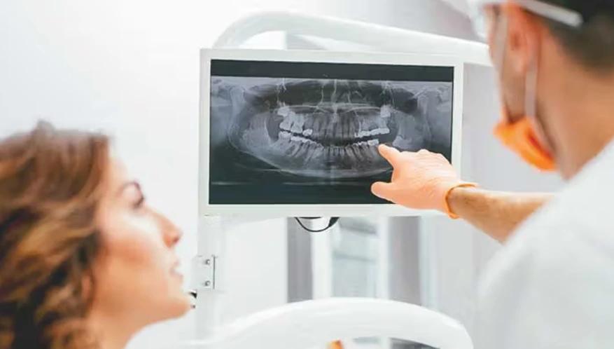
At Lema Dental Clinic, D0230 is done with digital radiography, which is more comfortable for the patients and exposes them to less radiation. According to Dentist Polen Akkılıç, digital sensors come instead of traditional film, thus the pictures are taken immediately and shown on the monitors right there. This modern way not only makes the diagnosis more precise but also makes the patients more comfortable and aware.

Use of D0230 is a must if the doctor wants to see as many different areas as possible to understand the ailment better or to follow up with the healing after the surgery or just to be sure of the health of neighboring teeth before insertion of an implant. In that case, it is fully covering and clinically confirming the diagnostic requirements.
When is Dental Code D0230 Used?
D0230 is employed for additional periapical images if different teeth or areas require investigation. That is, after taking the very first periapical X-ray (D0220), more films may be necessary to get a clear view of the adjacent parts that are sensitive, mobile, or have visible lesions.
Professor Doctor Coşkun Yıldız says that this code at Lema Dental Clinic is mainly involved in situations such as:
- Root canal therapy (endodontic treatments) where the condition of multiple teeth in one quadrant is required
- Planning of dental implant and control after the surgery
- Identifying caries under the restorations
- Checking bone levels around several teeth
The point of the idea for the additional films is always the requirement in the clinic, thus the use of radiographs is done ethically. D0230 is a tool that helps to achieve diagnostic completeness without the need for excessive imaging, which ensures both correctness and patient protection.
The Role of D0230 in Modern Dentistry
Correct diagnosis is the lifeblood of modern dentistry. D0230 act as a crucial supplement to the initial X-ray that can essentially be considered a basic sketch of patient’s oral structures. With the help of advanced technology like cone-beam computed tomography (CBCT) or panoramic radiography, periapical films come closest in providing ultra-fine details in locating anomalies on a micro-level.
Dentist Polen Akkılıç points out that D0230 is indispensably significant in the field of endodontics, whereby the determination of success is the detection of area of abscess or minute root fractures. Missing one single lesion may result in pain, infection, or retreatment. Through utilization of additional periapical images, the clinician guarantees that no root canal or the surrounding tissue is left uninspected before proceeding.
At Lema Dental Clinic, the process of imaging is supported by AI-assisted diagnostic software which makes the invisible obvious. Every shot made under D0230 is saved digitally in the clinic’s record system, which is a great tool for long-term patient progress monitoring over different visits.
How D0230 Is Performed at Lema Dental Clinic
At Lema Dental Clinic, the performance of every D0230 operation is in line with rigorous safety and quality standards. Before going for more X-rays, Dentist Polen Akkılıç and her team assess the need for it and check the patient’s history to make sure the radiation exposure will be at a minimum.
The step is initiated through the insertion of a digital sensor into the oral cavity that is aligned with the targeted tooth area to obtain the X-ray. The doctor directs the X-ray machine to the right angle, thus allowing for a complete view of the root apex and its surrounding bone. The place where the image is stored is ready straight after the capture, enabling both the doctor and patient to have a look at the results at the same time.
According to Professor Doctor Coşkun Yıldız, the current digital setups at Lema Dental Clinic use about 10% of the energy needed for traditional film-based X-rays, thus they emit drastically lower radiation. Furthermore, the institution provides their patients with lead aprons and thyroid shields. These measures make sure that every D0230 exposure is not only safe but also necessary from a diagnostic point of view.
What Are the Benefits of Using Dental Code D0230?
D0230 is a very instrumental code in implementing different clinical and administrative strategies for dentists and patients. The journey from accurate diagnosis to proper documentation would not be complete without the intervention of this code.
Key advantages of D0230 are:
- Better visualization of dental and bone structures
- At early stages discovering the infections, fractures, and root resorption
- Precise observation of the results of endodontic and implant procedures
- Elimination of diagnostic doubts during treatment planning
- Provision of legal and insurance documentation through precise coding
At Lema Dental Clinic, the image that goes with D0230 is the main source of a clearer diagnostic foundation. Dentist Polen Akkılıç notes that these X-rays often reveal conditions that might otherwise go unnoticed in panoramic imaging. Thus, by recognizing the initial symptoms of the disease, dentists become capable of providing targeted and less invasive care.
Warnings and Precautions About Dental Code D0230
Even though dental X-rays feature a very low radiation dose, the safety of patients is still the foremost concern. To minimize cumulative exposure, the Professor Doctor Coşkun Yıldız is recommending the very moment the “ALARA” principle should be strictly followed for D0230, i.e., As Low As Reasonably Achievable.
Before going for any radiographic procedure, pregnant patients and people who are sensitive to radiation should definitely inform their dentist. International radiation safety standards are followed by Lema Dental Clinic ensuring that all patients use protective gear and lead aprons.
Precise and safe are ensured through regular upkeep of the radiographic machines, their timely calibration, and strict adherence to exposure guidelines. The clinic’s digital system will automatically lower or raise the radiographic exposure based on patient age and size, thus further reducing radiation risk.
When Should You Avoid Using D0230 and What Should You Use Instead?
Cases which lead to the same diagnostic conclusion with less number of exposures should not use D0230. If there is a need for a full-mouth examination, a panoramic X-ray (D0330) or cone-beam CT (D0367) might be a better choice. Both of them offer a wider anatomical view in just one scan.
Among other things, Lema Dental Clinic‘s Dentist Polen Akkılıç and Professor Doctor Coşkun Yıldız make their decision on the best imaging method based on the complexity of each case. They often resort to D0272 (bitewing, two films) or D0274 (four films) for their young patients in order to minimize radiation exposure while still obtaining sufficient diagnostic information.
If soft tissue or joint evaluation is required, CBCT or MRI can be used as a supplement to D0230. The main goal is always to keep a balance between the diagnostic benefit and patient’s safety and comfort.
Case Study for D0230
A 46-year-old male patient came to Lema Dental Clinic complaining about the continuation of discomfort after root canal therapy. The first X-ray under D0220 didn’t show any clear pathology, but Professor Doctor Coşkun Yıldız decided to take additional films under D0230 to check the adjacent teeth. The second image pointed out a hidden lateral canal infection spreading to the bone.
Under the microscope, a small endodontic retreatment was done, and the patient’s pain disappeared after a week. Dentist Polen Akkılıç underlined that if the D0230 image was not there, the lesion might have stayed unidentified, thus resulting in chronic infection or tooth loss. This example demonstrates how crucial is the diagnostic value of additional periapical imaging.
During follow-up sessions, D0230 films were utilized to confirm the recovery and bone regrowth, thus highlighting the role of multi-angle diagnostics in achieving successful outcomes.
| Code | Description | Purpose | Image Count | Performed By |
|---|---|---|---|---|
| D0220 | Intraoral periapical, first film | Initial diagnostic image | 1 | Dentist Polen Akkılıç |
| D0230 | Intraoral periapical, each additional film | Additional diagnostic coverage | 2 or more | Professor Doctor Coşkun Yıldız |
| D0274 | Bitewing, four films | Cavity detection between teeth | 4 | Lema Dental Clinic hygiene team |
| D0330 | Panoramic radiographic image | Full mouth and jaw overview | 1 | Radiology department, Lema Dental Clinic |
References
- American Dental Association. (2024). CDT Code D0230: Intraoral Periapical X-Ray – Each Additional Film. Chicago, IL: ADA Publications.
- Yıldız, C., & Akkılıç, P. (2024). Digital Radiography and Diagnostic Imaging in Modern Dentistry. Istanbul: Lema Dental Clinic Research Journal.
- Farman, A. G., & Scarfe, W. C. (2023). Radiographic Techniques and Interpretation in Dentistry. Elsevier Health Sciences.
- American Academy of Oral and Maxillofacial Radiology. (2023). Radiation Safety and Patient Protection Guidelines.
- Langland, O. E., & Sippy, F. H. (2022). Principles of Dental Imaging (3rd ed.). Lippincott Williams & Wilkins.
Frequently Asked Questions About Dental Code D0230
It refers to each additional intraoral periapical X-ray beyond the first image taken during diagnosis.
They provide additional views to detect hidden infections, fractures, or decay not visible in the first image.
Yes, especially when performed with digital systems like those at Lema Dental Clinic, which use minimal radiation.
Each additional film takes about 2–3 minutes, depending on positioning.
Yes, it shows bone levels around the tooth roots, helping in periodontal diagnosis.
Only when clinically necessary—your dentist determines the frequency.
Dentist Polen Akkılıç and her radiology-trained team under Professor Doctor Coşkun Yıldız.
Yes, most dental insurance plans cover additional X-rays under preventive or diagnostic services.