

When you have a toothache that is lasting and you see that your gums are swollen, the very first thing that comes to your mind is “Do I have an infection?”. To confirm this, the dentist will probably advise a dental X-ray. This is a common step that is the basis of modern dental diagnostics; thus, it is a must-have tool in the fight for health beyond what is visible to the naked eye.

This ultimate manual informs you about the ways dental x-rays detect infections that have gone unnoticed, the different kinds of x-rays used, and the reason why this technology is necessary for saving your smile and keeping you healthy. Knowing about this operation helps you to be a part of your dental care and thus you can get the right diagnosis and treatment that is both prompt and efficient.
The Direct Answer: How Dental X-Rays Reveal Hidden Infections
A dental X-ray is an accurate and dependable method to determine the state of teeth and the jawbone. So, if there is any infection around the roots of the teeth or in the jawbone, it can be located with the help of a dental X-ray. Dental x-rays are detailed pictures of the hard structures in your mouth. Healthy bone and enamel coat of the tooth show up as bright areas on the X-ray film or sensor since they are dense and absorb a higher amount of the radiation. On the other hand, infections lead to certain changes in the images that a trained dentist can recognize instantly.
The most typical indication of an infection on a dental x-ray is a dark area that can be seen at the very point of the tooth root. This darkness stands for the loss of bone, the direct consequence of the body’s inflammatory reaction to microbial invasion. The particular kind of abscess that is called a periapical abscess is one that originates from the tooth and finally infects the area where the root apex meets the bone thus, it breaks down the bone around it. After spotting this clear radiographic marker, your dentist makes a confident diagnosis of this condition thus, he is able to locate the infection and determine its exact position.
Beyond the Root: The Types of Dental Infections X-Rays Detect
Dental x-rays are excellent tools in the diagnosis of four main types of oral infections that have different symptoms and health risks.
- Periapical Abscess: A periapical abscess is an infection that comes from the pulp of the tooth, which is the soft inner part with nerves and blood vessels. The most common cause of this situation is untreated tooth decay, where bacteria get in through a crack or a hole in the tooth, or as a result of an injury that makes the pulp infected and later on dead. The infection makes its way through the root canals and after that it exits the apex, thus, the formation of the typical dark shadow on the x-ray. The local infection that resulted from this can be controlled and stopped by detecting it early with the help of an X-ray.
- Periodontal Abscess: It is an infection that happens in the dental support structures, i.e., the gums and the alveolar bone. Usually a consequence of severe gum disease (periodontitis), a periodontal abscess on an X-ray looks like a dark, vertical wedge along the side of the tooth root. The most important point that this indicates is the very moment when the bone that is holding the tooth tightly is being destroyed. X-rays serve as a means to find out how far this bone loss has gone and to make the right plans for treatment to keep the teeth safe.
- Pericoronitis: A pericoronitis situation occurs when the area of the soft tissue next to a partially erupted tooth, usually a wisdom tooth, gets infected. Although the infected soft tissue might not always be clearly seen on an X-ray, the image is very helpful in showing the position, angle, and amount of space of the tooth. With this data, the dentist is able to figure out the actual cause of the infection and give the proper treatment suggestion.
Тhe Diagnostic Toolkit: Different X-Rays for Different Diagnostic Needs
To reveal the whole dental health picture, the dentists take various X-rays, each of which shows the area from a different angle.
- Bitewing X-rays: They show the upper and lower back teeth in one view. They are extremely good at cavity detection in between teeth and are used for checking if the fillings are still good. Although with their help, you can see very advanced bone loss due to gum disease, they are not the main instruments for finding infections at the root tips.
- Periapical X-rays: Such an X-ray zooms in on one or two full teeth, starting from the crown and going down to the root tip and the bone around it. It is the cleanest, closest, and direct means of pinpointing a periapical abscess, showing all the dental structures in detail, and clarifying the situation.
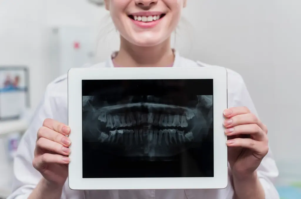
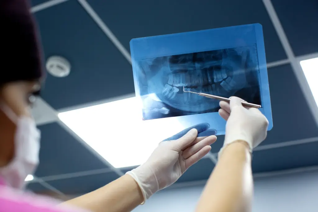
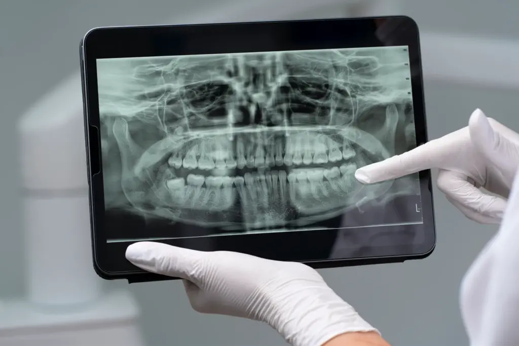
- Panoramic X-ray (Panorex): The device revolves around your head and thus, is able to make a single, broad image of all your teeth, jaws, temporomandibular joints (TMJs), and even sinuses. It provides an excellent overview and is consulted when assessing wisdom teeth, implant planning, finding cysts or tumors, and rooting checks for infections.
- Cone Beam Computed Tomography (CBCT): In complicated cases like complex root canal systems, dental implant planning, or continuous pain of unknown cause, the dentist uses a CBCT machine. This cutting-edge instrument creates detailed 3D pictures and thus, it gives the closest possible view of the bone framework, nerve paths, and the exact anatomy of an infection.
When Symptoms Signal the Need for an X-Ray
It is true that some dental infections can be silent at the beginning, but still, in most cases, your body gives you very obvious signs. If you get any of the following symptoms, then it is absolutely necessary to arrange a dental visit as soon as possible. The X-ray will definitely be one of the steps in figuring out the cause.
- Persistent Throbbing Toothache: An ongoing, very painful situation that can extend and affect the jawbone, neck, or ear is, without a doubt, the major sign of a pulp infection.
- Sensitivity to Hot and Cold: The sensitivity that lasts even after the hot or cold source has been removed is most likely coming from the nerve inside the tooth which is either damaged or dying.
- Tenderness When Chewing or Biting: The pain that occurs when you apply pressure to a certain tooth can be a sign of an infection at the root tip.
- Swollen, Red Gums: The swelling directly around one tooth which is very similar to a pimple, is the common symptom of an abscess.
- Facial or Cheek Swelling: The moment when the infection is beyond the immediate tooth area and thus, this swelling is the result, requires a visit to the dentist very urgently.
- A Bad Taste in Your Mouth or Persistent Bad Breath: These things can be a result of pus that is coming from an abscess and is being released into your mouth.
Safety First: Understanding the Minimal Risks of Dental X-Rays
Reasons for radiation-related anxiety are understandable. Patients should be aware, that the radiation dose from modern dental X-rays is very low. Safety measures put in place by dental practitioners are there to reassure patients of their safety.
- Digital X-ray Technology: Most dental clinics have already adopted the use of digital X-rays, which involve less radiation and, therefore, exposure to radiation is decreased by 90% compared to the old-fashioned film X-rays.
- Focused Beam and High Speed: The X-ray beam is tightly collimated so only the area that is of interest is being focused on and the exposure time is just a few seconds.
- Lead Apron Shielding: The application of a lead apron with a thyroid collar is a normal safety measure which, in effect, protects the rest of your body from any radiation that it might be coming from the sides.
The diagnostic benefit of dental X-rays is so great that the small risk that goes with it is barely noticeable. The health risk of leaving an infection undiscovered and unaddressed is much bigger than the one from exposure to radiation during an X-ray.
What an X-Ray Cannot Show: The Limits of Radiographic Imaging
Despite the power of dental X-rays, they are a part of a full diagnostic process and not the only one. There are also some restrictions on what they are able to uncover.
- Early-Stage Infections: Only in the initial stages of a pulp infection, the bone close to the root tip may not have loosened so much as to give a dark spot that can be seen on the x-ray. The dentist will determine the diagnosis based on your symptoms and tests.
- Soft Tissue Infections: X-rays depict hard tissues. An infection that is confined only to the gum tissue (gingiva) and not the bone may not be directly visible on a standard x-ray. The dentist will diagnose this during his/her examination by looking and touching.
- The Exact Bacterial Strain: An X-ray is used for locating the infection and provides an indication of the involvement but doesn’t reveal bacterial strain. However, the identification of bacteria is generally not required for efficient treatment in most cases.
The dentist always combines the findings of the X-rays with a thorough clinical examination which consists of visual inspection, palpation, and percussion tests to arrive at a final, accurate diagnosis.
The Confident Path Forward: Diagnosis and Effective Treatment
When a dental issue is proved by means of a dental X-ray, the next step is treatment along the same logic which is clear, swift, and designed to achieve the three goals: dissipate the infection, take away the pain, and bring back the health.
- Root Canal Therapy: In the case of a tooth with a periapical abscess, the standard solution is root canal therapy. This treatment involves the removal of the infected pulp inside the tooth, the sanitation and disinfection of the inner spaces, and then closing the space with a material that is both secure and compatible with the body. Afterward, the tooth is covered with a crown that will serve two purposes – protection and restoration of the complete function. This treatment keeps great success for a very long time and thus, you are able to retain your natural tooth.
- Tooth Extraction: In the case that the structure of the tooth is extremely damaged and thus, saving is not an option or the infection is very severe, removal may be the most sensible way to go to keep the rest of the bone and tissues healthy. Your dentist will talk about the options for replacing the tooth, e.g., a dental implant or bridge, to be able to keep the smile’s alignment and functionality.
- Periodontal Treatment: The treatment for a gum disease infection usually, comprises undergoing scaling and root planing, a deep cleaning method. This way, the procedure not only cleanses the plaque and tartar that have formed below the gumline but also removes the roughness from the roots of the teeth, thus, helping gums to attach again. If the case is severe, then a specialist in periodontics might do some surgical operations aiming at producing more bone and tissue that have been lost.
- Antibiotic Therapy: Antibiotics help to ease the symptoms of dental infections and should not be seen as a cure. If the dentist sees that an infection is spreading over the face and causing swelling and other systemic symptoms like fever, then only he will decide to prescribe them. Nevertheless, the one that brings about the solution will always be the one that actually deals with the source of the problem interventionally, e.g., root canal or removal.
Your Role in a Healthy, Infection-Free Smile
The best remedy is prevention. You can take an active part in significantly lowering the chances of getting dental infections by keeping a good and consistent oral hygiene routine that is consistent.
- Brush Twice Daily: A fluoride toothpaste together with a soft-bristled toothbrush should be used, and the brushing should last for two minutes every time.
- Floss Every Day: Flossing is an absolute must if one wants to get rid of plaque and food remains that are in between teeth and thus cannot be reached by a brush.
- Have Regular Dental Check-ups: In six-month intervals, in addition to the cleanings that are done by the professionals, it is very important to have the check-ups because they take care of the removal of tartar that has become hard and, also, they are in time for spotting small cavities or early stages of gum disease before they have a chance to become serious infections.
- Keep a Balanced Diet: In addition, it is better to eat less sugar and acidic products and to drink less acidic drinks, which are the main contributors to the decay process.
- Drink Enough Water: A large amount of water consumption helps the mouth to rid itself of leftover food and bacteria and is also very good for saliva, the mouth’s natural defense system.
A Vital Tool for Your Health

Without a doubt, the answer to the question “Can a dental X-ray show an infection?” is yes. One of the safest, most reliable, and indispensable means for diagnostics is dental X-ray, which shows the hidden area to the dentist, helps him figure out the source of the problem correctly, and, finally, comes up with a precise treatment plan.
Apart from that, to mention the highest value of X-rays, it is their ability to shed light on very early-stage hidden infections, and thus, dentists and patients get enough time to take proactive and effective measures against them in order to relieve pain, save natural teeth, and secure oral and general health for a long time to come. One of the cornerstones of getting the confident, high-quality dental care that you deserve is trust in this technology.
Sources:
- White, S. C., & Pharoah, M. J. (2014). Oral Radiology: Principles and Interpretation (7th ed.). Mosby.
- American Association of Endodontists (AAE). (2023). Signs and Symptoms of Endodontic Disease.
- American Academy of Periodontology (AAP). (2023). Gum Disease Information. //www.perio.org/for-patients/gum-disease-information/
- MedlinePlus. (2022). Pericoronitis. U.S. National Library of Medicine. //medlineplus.gov/ency/article/001967.htm
- Food and Drug Administration (FDA). (2022).Dental X-Rays.
- Carter, L., & Farman, A. G. (2008). Panoramic Radiology: A Review. Seminars in Orthodontics.
- Scarfe, W. C., & Farman, A. G. (2008). What is Cone-Beam CT and How Does it Work? Dental Clinics of North America.
Frequently Asked Questions About Dental X-rays and Infections
It is indeed possible for immunologists of medical imaging immunologists to see hidden infections on dental radiographs. What is more, for example, abscesses and periodontitis can be detected from the X-ray pictures even before they can be felt or seen by the naked eye. Therefore, the physician can practice intervention at an early stage, and the efficiency of the treatment will be considerably higher.
Usually, the decision about dental X-rays is made by the doctor and it varies according to one’s current oral health, age, and risk profile. As a rule, X-rays are recommended once every two years for the adult population; However, if one has ongoing dental problems, then he/she need to see the dentist more often.
Yes, dental X-rays are very safe. The radiation exposure is minimal, and dental professionals take steps to ensure your safety. If you are pregnant, be sure to inform your dentist.
The answer is yes. Along with infections, dental X-rays can show dental caries (cavities), bone loss (the process of the bones becoming frail), tumors, cysts, and other mouth problems that may not be seen or felt.
The time interval between an infection and its appearance on an X-ray varies greatly depending on the degree of infection. For instance, abscesses are infections that can be very easily and quickly located, while the visibility of other infections in an X-ray might be delayed for several weeks.