

Can a Dental X-Ray Cause Cancer? A Comprehensive and Reassuring Guide to Safety and Benefits
When the dentist wants to take an X-ray of your teeth, you naturally start wondering. These days, where there is plenty of health information, concerns about radiation and cancer after some years are still common and understandable. You deserve to get answers that are clear, accurate, and based on science so that you can make decisions about your healthcare with confidence. This comprehensive guide is your support when you have no clue what is going on and the fact that you are nervous. We will delve into the tightly guarded safety features of the current dental X-rays, talk about their pleasant side – the diagnostic benefits, and, to comfort you, we will bring the facts that dental X-ray is a safe and lifesaving tool.
Our main point during the whole discussion is your health, and we want to help you get the information that will make you feel safe and be able to make up your mind during your dental care.
Are Dental X-Rays Safe? A Resounding Yes, Backed by Rigorous Science
The categorical answer is affirmative; present-day dental X-rays are a very safe and well-controlled diagnostic measure. All dental radiology is based on a core international safety principle called ALARA, which means “As Low As Reasonably Achievable.” It signifies that the dental team uses the lowest dose of radiation which is still able to provide a diagnostically useful image.
- Technological Advancements: The transformation from conventional films to digital X-ray sensors is an enormous safety improvement. Digital radiography uses much less radiation – up to 90% less – than that of the traditional film-based systems in order to get the same quality of the image. This technology makes it possible for the dentist to obtain a perfect image in an exposure time of only a few milliseconds.
- Precision Beam Collimation: The X-ray device along with the collimator doesn’t emit a broad beam of rays. On the contrary, it utilizes a collimator, a device that narrows down and focuses the beam into the most exact spot on a tooth or a small area of the jaw. This focus makes sure that the closest tissues get the least possible amount of radiation.
- Mandatory Protective Shielding: The lead apron and thyroid collar are not only accessories; they are part of the protocol. Lead is a heavy metal and can hardly be penetrated by radiation. Putting this apron over the chest and the collar around the neck ensures complete protection of the vital organs and the thyroid gland against even the tiniest amount of scattered radiation.
- Professional Expertise and Licensing: Dental professionals are taught in detail about the safety of radiology during their educational program. Besides that, they learn the right methods of positioning the machine, the patient, and the film/sensor to achieve the best results with the least possible exposure. State regulatory bodies also check dental offices from time to time to make sure that all the devices are working properly and safely.
If you add to that the cutting-edge technologies and the strict rules for safety, a dental radiograph is a low risk procedure which you can consider as a routine and its benefits are vital for your oral health.
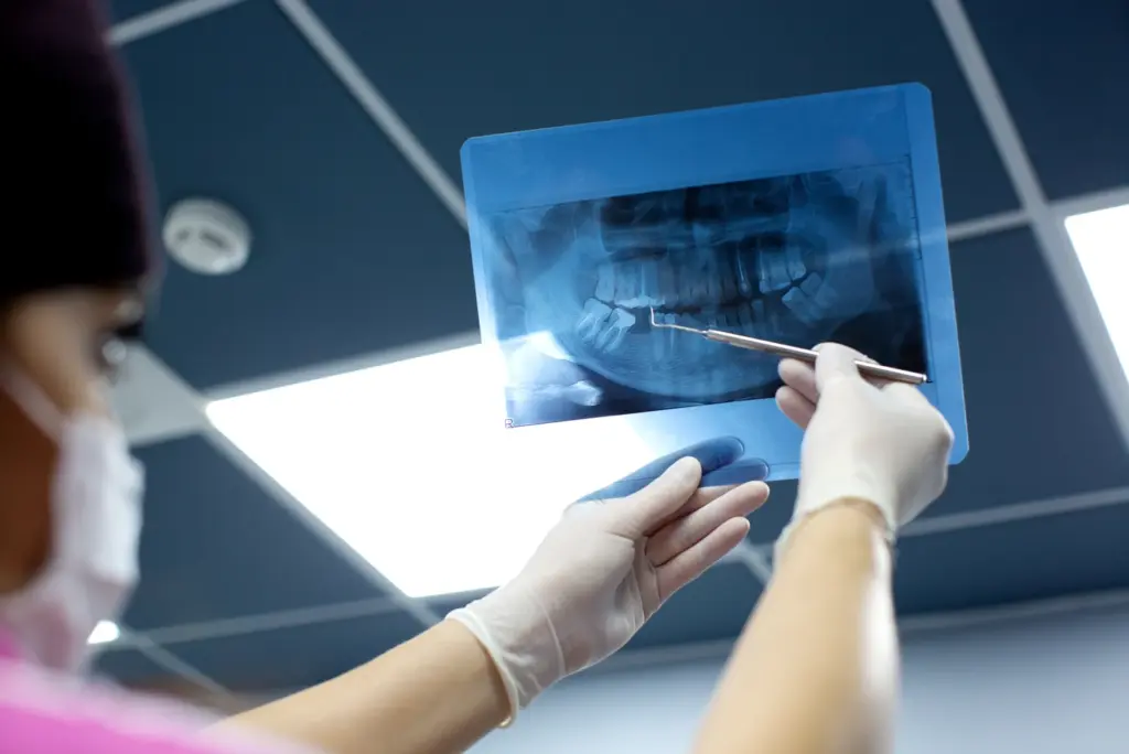
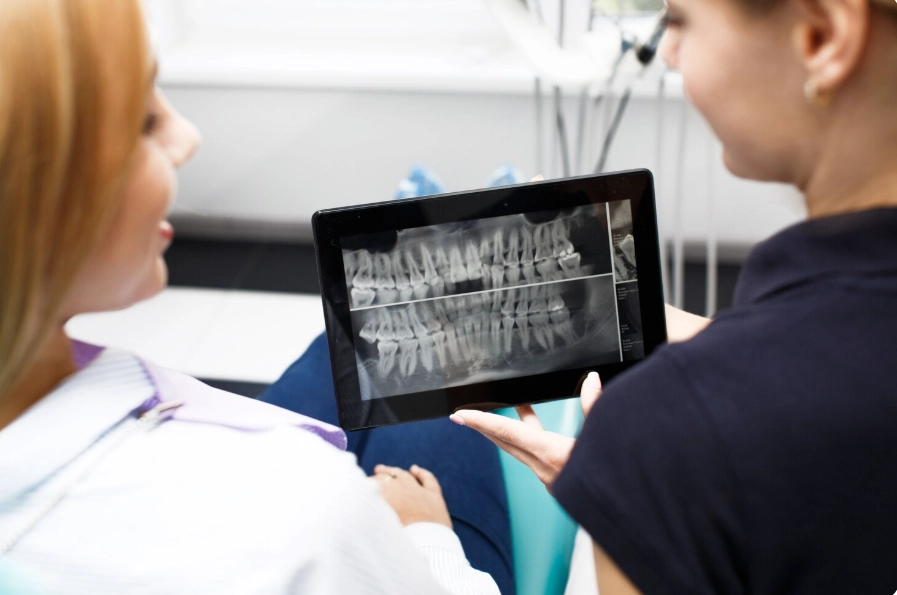
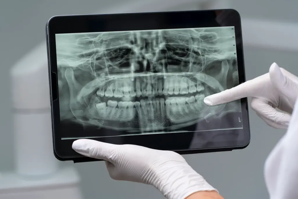
What Does a Dental X-Ray Show? The Unseen World Beneath the Surface
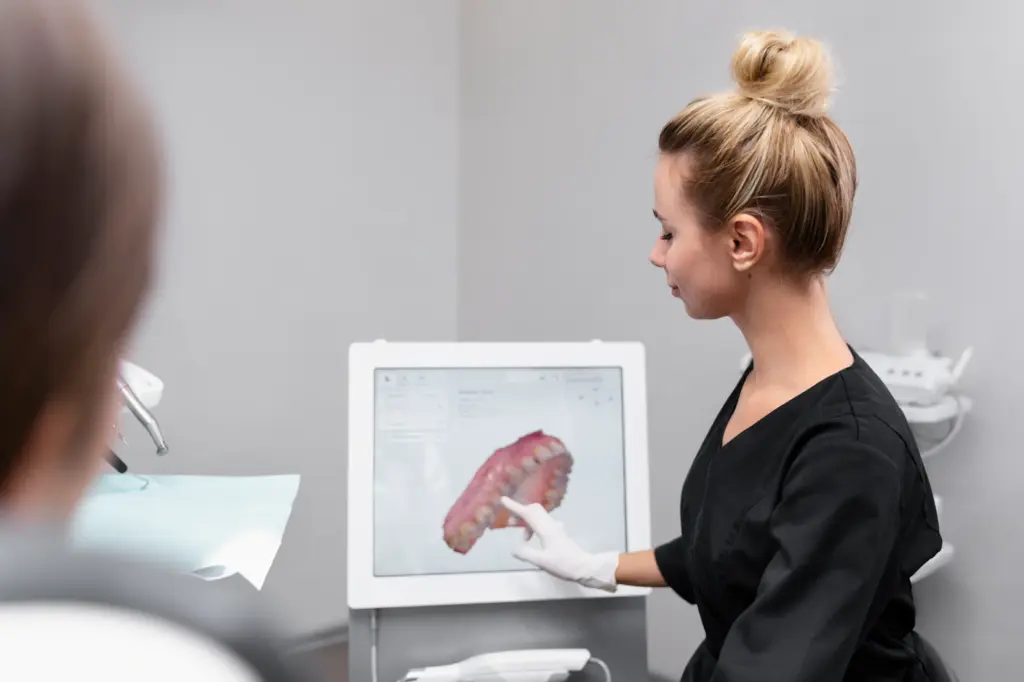
Visual inspection is limited in what it can do. It helps the dentist see only the outer parts of the teeth and gums, but the major portion of the mouth’s structure is still a secret to him. Dental radiography is an essential guide that leads the way to the parts under the surface and in between the teeth. With this, the dentist can make a leap from speculation into precise and final diagnosis. These images hold a treasure of information which is integral for the complete treatment:
- Early-Stage Decay Between Teeth: Most of to decay occurs in the side surfaces between two teeth where a mirror and explorer can’t reach. Dental X-rays uncover these parts that have lost minerals long before they become big enough for a source of pain or requiring extensive treatment, thus allowing for the least invasive intervention.
- Precise Assessment of Bone Loss: Periodontal (gum) disease is the process that leads to the destruction of the jawbone, which supports the teeth. Through X-rays, one can get very accurate bone level measurements and hence, determine the stage of gum disease, track its progression, and evaluate the effectiveness of therapy. This plays an important role in the prevention of tooth loss.
- Identification of Impacted Teeth: The third molars sometimes the canines, may be enclosed (impacted) inside the jawbone. X-rays depict the exact location, direction, and developmental stage of the impacted teeth as well as their closeness to the nerve canals and next teeth, which is indispensable in the process of safely planning.
- Detection of Abscesses and Infections: X-ray pictures of a periapical abscess i.e. at the root tip of a tooth, or a periodontal abscess in the gum, will show it clearly. These infections may be silent, without any noticeable symptoms, but they can cause bone damage and spread to other areas of the body if they are not treated.
- Revelation of Cysts and Tumors: Dental X-rays can spot abnormal growth in the jawbones, like cysts (sacs filled with fluid) and tumors – both benign and malignant. Finding these issues early, including oral cancer that originates in the bone, can save a person’s life.
- Evaluation of Dental Restorations: X-rays make it possible for your dentist to evaluate the condition of the restorations that have been done. They can check if there is a gap where decay can get in again in a filling, if a crown fits perfectly, or if there is any hidden damage to a tooth which has been through a root canal.
Dental X-rays, by supplying such a detailed look inside, give your dentist the power to draw up a preventive, tailored, and effective treatment plan that deals with problems at their earliest, most controllable stages.
Are Dental X-Ray Images Really Necessary? The Critical Role of Proactive Diagnosis
It is a clue that a patient is engaged and responsible when they question the necessity of a medical procedure. In the case of dental X-rays, they are indeed an indispensable part of a proper and comprehensive oral health check-up. Not having an X-ray is like an instruction to your dentist to provide care with incomplete information which is similar to a pilot flying without seeing. Hidden dental issues may be the ones that cause the biggest trouble later on because, in most cases, such problems do not show any symptoms and only become painful after they reach the advanced stage.
Think about these examples in which an X-ray is not only helpful but also necessary:
- A person without any pain could have a huge decay between two molars that has not yet been able to affect the nerve.
- An individual with well-groomed gums might have a considerable loss of bone underneath the gumline, which is the first manifestation of severe periodontitis.
- One might have a small, non-painful abscessed area at the root of a tooth that was given a root canal a few years ago and which may lead to a serious systemic infection over time.
The American Dental Association sets very clear rules about how often you should have X-rays-voluntarily depending on your health condition- age, risk of diseases, symptoms, and medical history are the factors taken into consideration. The tiny, precise amount of radiation that is used in one single X-ray is very far from being a deterrent when you weigh it against the incredible early detection and prevention benefits. The possible outcome of not uncovering a hidden issue such as a complicated infection, a growing cyst, or early-stage oral cancer is way beyond the very slight radiation exposure that is only a theoretical risk. Therefore, it would be a very smart, proactive and courageous move to accept the schedule of X-rays advised by your dentist when it comes to securing not only your oral health but also your general well-being of body.
Dental X-Ray: How Much Radiation? Putting Minuscule Numbers into Real-World Context
It’s very important to know the real amount of radiation that is involved if you want to be able to calm your worries off. The exposure to radiation is recorded in units called microsieverts (µSv). To give accurate and useful information, let us look at the radiation from normal dental X-rays against that from regular background and other typical activities:
- Four Bitewing X-Rays (Digital): A local check-up series, which is used for the detection of interproximal caries in the posterior teeth, generates an effective dose of around 5 µSv.
- Panoramic X-Ray (Digital): In this case, one picture that shows the whole jaw along with the teeth results in about 10-25 µSv.
Let’s see these figures using the real world:
- Around 8000 µSv of background radiation are inhaled per year by an average American due to the nature of the environment. The average daily exposure comes to about 22 µSv.
- The amount of radiation for a series of four bitewing X-rays (5 µSv) is almost one single day of natural background exposure.
- A panoramic X-ray (25 µSv) has the same amount of radiation as what a human being would absorb for about three days just living on earth.
- Some more helpful contrasts: Flying on a plane cross country from New York to Los Angeles will expose a passenger to about 40 µSv of extra cosmic radiation due to the altitude. This is quite a bit more than what you would receive during a bitewing series.An individual residing in a stone, brick or concrete house for a year will add roughly 70 µSv to his/her natural radon gas exposure. The use of tobacco is a far more significant contributor to the atomization of lungs and, thus, smokers are subjected to around 160,000 µSv annually.
- Flying on a plane cross-country from New York to Los Angeles will expose a passenger to about 40 µSv of extra cosmic radiation due to the altitude. This is quite a bit more than what you would receive during a bitewing series.
- Living in a stone, brick, or concrete house for a year adds about 70 µSv due to natural radon gas.
- Using tobacco is a far more significant source of radiation exposure to the lungs, with smokers receiving an estimated 160,000 µSv per year.
All of these comparisons point to one thing only – the radiation exposure arising from dental X-rays for diagnostic purposes is just a tiny addition to the natural background radiation that we are always subject to.
Does Dental X-Ray Radiation Cause Cancer?
The notion that even a small amount of radiation may lead to cancer derives mainly from a model which assumes that there is a linear relationship between the dose and the effect without any safe threshold, and thus it is based on very high doses, such as those encountered by the survivors of atomic bombs. In contrast, the risk associated with extremely low doses employed in dental imaging is so minute that it is practically negligible – very close to zero – according to several international organizations such as the American Cancer Society and the National Cancer Institute.
Understanding this comes down to the idea of risk-benefit assessment. Besides being extremely small, the radiation dose from a dental X-ray is also very limited to the most radiation-resistant parts of the body, i.e., teeth and jawbone. The estimated theoretical risk of causing a fatal cancer from a full-mouth series of X-rays is somewhere of the order of 1 out of 2 million. To compare this almost unfathomably tiny figure with something more familiar, your life-time luck of being hit by lightning is about 1 in 15,000.
The company of this almost immeasurable theoretical risk with the very real, verified, and substantial advantages of dental X-ray goes as follows:
- They reveal what is truly happening inside the body in the case of decay, infection, and bone loss.
- By allowing early-stage cancer detection, e.g. oral cancers originating in the jawbone, they act as life savers. An X-ray can actually save your life by finding cancer at an early stage.
- By identifying issues long before they become symptomatic, they avert pain and complex emergencies.
- With the help of X-rays, treatments become fruitful and you get faster healing, e.g. root canals, implants, and extractions.
When you put the almost non-existent hypothetical risk side by side with the huge, real benefits of the accurate diagnosis and disease prevention at early stages, the worth of dental X-rays becomes very clear, in fact, overwhelming. They are not a source of worry but, actually, a very powerful and necessary means of securing your future health.
Can Dental X-Rays Cause Thyroid Cancer?
This question was one of the most serious ones in the past. Even so, it is now answered, and dental specialists deal with the situation in a totally different way. Large population studies failed to detect causality between new-generation dental X-rays and the increased incidence of thyroid cancer. Mainly it is because two reasons are responsible, one being the extremely low radiation dose and, most notably, the lead thyroid collar’s usage is almost always universal.
The thyroid gland located in the neck is the most radiation-sensitive one among the rest of the body tissues. It is well aware that dental safety postal regulations dictate that in case of any intraoral X-ray (where the film or sensor is put in the mouth), a lead thyroid collar should be fitly placed over the patient’s neck. The collar is not a sign; rather, it is a very effective physical protection. A lead lining is an absolute barrier to the scattered radiation that might be coming from the primary beam; thus, the thyroid gland gets zero irradiation.
On one side, the ultra-low radiation digital technology of today and on the other, this obligatory, efficacious protective shield – as a result, the risk of the thyroid gland from a properly done dental X-ray is non-existent. Your dental team will certainly not miss this step if they are well-trained to do it for every single patient, for every single X-ray, making the safety of the thyroid a care standard that cannot be compromised.
Are Dental X-Rays Safe During Pregnancy?

During pregnancy, oral hygiene should be maintained at the highest level, which is perfectly safe and, in fact, highly recommended by both dental and medical professionals. The hormonal changes may cause pregnancy gingivitis and can lead to other oral health problems; all these have been linked to negative pregnancy outcomes such as premature delivery and low birth weight. So, dental problems solving becomes a very important part of prenatal care.
The two leading professional bodies supporting the view are American College of Obstetricians and Gynecologists (ACOG) and the American Dental Association (ADA). They jointly confirm the safety of dental X- rays during pregnancy. The radiation emitted during a dental X-ray is at a very low level and is focused on the mouth area, rather than the abdomen. Carrying out the procedure under a lead apron with a thyroid collar offers double protection, thus, impeding the penetration of radiation to both the mother’s torso and the fetus.
On the condition that you are a carrier of an unborn baby and it is not your usual yet, then it is always wise to reschedule your routine X-ray right after giving birth in a purely conservative way. Even so, it might be so hard to stand the pain without seeing the source if you got into a dental emergency such as severe toothache, swelling, or mouth injury. In such an event, not only will it be safe but it will also be a medical necessity to take the X-ray. In fact, an accurate diagnosis is the only way to the effective treatment, which eventually leads to the eradication of the infection source and the reduction of the stress factor, both of which could be far more harmful for the pregnancy than the X-ray itself. Dentists are always pleased to take into consideration your existing condition during your check-up and thus, they can take all the care steps needed; however, you can be sure that taking an X-ray when necessary is not something that can cause harm to your baby.
Dental X-Ray Before I Knew I Was Pregnant: Complete Reassurance for an Early Exposure
It can’t be said that it was not the best of times for an early pregnancy period but a dental X-ray went on without your knowledge. Now, you have every reason to be relieved. The amount of radiation given off by one dental X-ray is very limited and localized; thus, it is not considered a risk to the embryo at its earliest stages of development. The radiation dose is way below the minimum level threshold that could lead to developmental problems.
Informing your dentist and obstetrician about the incident is always something good to do and then they can record it in your medical history. Nevertheless, they will most definitely concur that there is no reason for fear or any need for follow-up monitoring concerning the X-ray. The great advantage of having your oral health condition diagnosed and treated at that time was far more than enough to compensate for the tiny risk. With the passage of time in your pregnancy, your dental team members will keep on doing their work at the highest level, which is using the lead apron and thyroid collar whenever an X-ray is to be taken.
Sources:
- American Dental Association (ADA).The Use of Dental Radiographs: Update and Recommendations.Journal of the American Dental Association, 2019.
- Relevance:This is a foundational document outlining the official guidelines for dental X-ray use, frequency, and safety protocols, forming the basis for professional standards in the United States.
- American College of Obstetricians and Gynecologists (ACOG).Committee Opinion No. 569: Oral Health Care During Pregnancy and Through the Lifespan.2022 (Reaffirmed 2024).
- Relevance:This opinion, developed in collaboration with the ADA, provides authoritative confirmation that dental X-rays are safe during pregnancy with standard precautions, directly supporting the sections on pregnancy.
- White, S. C., & Pharoah, M. J.Oral Radiology: Principles and Interpretation, 8th Edition.Elsevier, 2018.
- Relevance:This is a leading textbook for dental and radiology students. It provides the detailed technical data on radiation doses (µSv) for different types of X-rays and the scientific principles behind digital radiography, collimation, and the ALARA principle.
- National Council on Radiation Protection and Measurements (NCRP).Report No. 184: Medical Radiation Exposure of Patients in the United States.2019.
- Relevance:This report provides comprehensive data on population radiation exposure, including from medical and dental sources. It helps contextualize dental X-ray doses against background radiation and other medical procedures.
- U.S. Food and Drug Administration (FDA).Dental X-Rays.[Webpage, Updated Feb 2023].
- Relevance:As the federal agency regulating medical devices, the FDA provides patient-friendly information on the benefits and risks of dental X-rays, emphasizing their safety and the importance of discussing needs with a dentist.
- American Cancer Society (ACS).Radiation Exposure and Cancer.[Webpage, Updated Dec 2022].
- Relevance:The ACS provides clear information on the difference between high-dose and low-dose radiation, stating that the risk from very low-dose exposures, like those in dental X-rays, is so small it is often undetectable.
- Brenner, D. J., & Hall, E. J.Cancer Risks Attributable to Low Doses of Ionizing Radiation: Assessing What We Really Know.Proceedings of the National Academy of Sciences, 2003.
- Relevance:This seminal paper discusses the challenges of extrapolating cancer risk from high to very low doses of radiation, supporting the concept that the risk from dental X-rays is theoretical and negligible.
FAQ’s: Can a Dental X-Ray Cause Cancer?
Yes. Dental X-rays use a tightly focused beam and significantly less radiation, targeting only your jaw. Medical X-rays for larger body areas require a wider beam and higher dose.
The risk is negligible. The extremely low radiation dose is considered safe, and the health benefits of early disease detection far outweigh any theoretical risk.
No. With modern digital technology and standard safety precautions like lead aprons, dental X-rays are not harmful and are a vital diagnostic tool.
No. The term “dangerous” does not apply. Stringent safety protocols make dental X-rays one of the safest medical imaging procedures available.
Yes. Dental X-rays can reveal signs of abnormalities, including tumors in the jawbone, playing a key role in early detection.