

Check texture: sticky or smooth?
It starts with a glance in the mirror. Suddenly, you spot a small dark stain on your molar or a yellowish shade near your gum line. Immediately, you feel a wave of panic. Is this too much coffee, or is my tooth already dying?
At Lema Dental Clinic, our clinical experience shows that this is the most common fear among our patients. We’ve had patients flying in from various parts of Europe, utterly convinced that their entire teeth need to be replaced with implants, only to be told that they just require a deep cleaning. On the other hand, we also have people who downplay their condition by saying they just have “a little stain”, while they are actually covering up a huge decay problem.
The distinction between a stain and a cavity (rot) is the distinction between a dirty window and a broken one. One is merely a matter of appearance; the other is a matter of structure.
Before you come to our dental clinic in Turkey, here’s a simple rundown on how you can figure it out yourself.
The “Sticky” Test: How to Tell by Touch
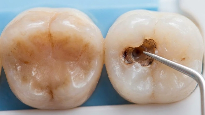
Pictures aren’t always a reliable means of identification. A dark brown spot may be just a coffee stain, while a white, chalky spot is actually a sign of severe decay. Professor Doctor Coşkun Yıldız is a great believer in the idea that one should not simply judge by color but also pay attention to texture.
- Stains are smooth: When you run your tongue or a dental explorer over a stain, it most likely has the same texture as the rest of your tooth—smooth, glass-like, and intact. It is merely color on enamel.
- Decayed/Rot/Cavity is sticky or rough: Tooth decay is the process of dissolving the minerals that make up the structure of the tooth. If the area feels rough, you can see depressions, or if a dental instrument slightly “sticks” in the spot, you cannot call it a stain anymore. That is a tooth cavity (caries). Enamel has caved in, thus a tiny hole has been formed at the location.
The Color Spectrum: What Your Tooth Is Telling You
Even though texture comes first, color certainly gives us some first-hand information.
1. Brown or Black Spots
- Stain: Stain basically signifies general yellowing or brown tint, most likely at the gum line or the inner surface of the bottom teeth. The causes are mostly smoking, coffee, or tea.
- Rot: This is the kind of damage that typically as a black or dark brown spot in one place. It is more like a hole or shadow beneath the surface of enamel, not just on it.
2. White Spots
- Stain: White stains hardly ever exist.
- Rot: This one kind of tricks you. In fact, a cavity starts as a chalky white spot (demineralization). You see the enamel loses calcium before it gets black and breaks. If you notice chalky whitish spots, then your tooth is in jeopardy.
Why “Wait and See” is Dangerous
The fault of the “only stain” theory is that decay happens silently. Usually, a cavity does not get painful until the nerve is involved (the pulp). Dentist Polen Akkılıç and her team put a lot of emphasis on early discovery since finding the “rot” in the white-spot phase frequently allows us to do the healing with fluoride or a minor treatment, instead of having to do a root canal.
Comparison: Is it a Stain or Decay?
In order for you to be able to judge your own smile, here is a list of various symptoms that we use to identify different problems here at Lema Dental Clinic.
| Feature | Surface Stain (Extrinsic) | Tooth Decay (Cavity/Rot) |
| Texture | Smooth, glass-like | Rough, pitted, or “sticky” |
| Sensation | Painless | Sensitive to sugar, hot, or cold |
| Location | Widespread (entire smile) | Localized (specific spot/crevice) |
| Cleaning | Can be scraped off/polished | Cannot be brushed away |
| Structural Integrity | Tooth remains strong | Tooth may chip or break |
| Treatment | Professional Cleaning/Whitening | Filling, Root Canal, or Crown |
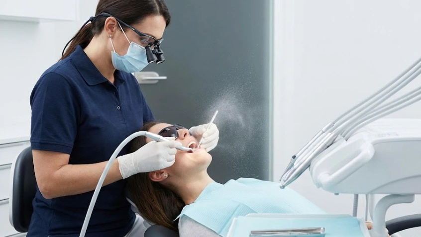
The Solution: How We Treat Both in Turkey
When you have a seat in a dental chair at Lema Dental Clinic, all the guessing ends. We employ an intraoral camera to display to you on a 4K screen exactly what is going on. After that, we know exactly how to proceed.
Vital Questions on Spotting Decay vs. Discoloration
Yes, but only in the very first stage. When decay appears as a “chalky white spot” (demineralization), it means the enamel is soft but not broken. At this stage, rigorous hygiene and high-fluoride treatments can harden the enamel again. Once the surface breaks (creates a hole), it cannot heal itself and must be filled.
It shouldn’t. If a “stained” area is sensitive to cold, heat, or sweets, it is not a stain at all, but leakage. This could be a cavity underneath an old filling or decay hiding between teeth where you can’t see it. Sensitivity is your nerves’ way of screaming for help.
No, and they can be dangerous. If you apply bleaching gel to a tooth with an active cavity (rot), the chemicals can penetrate through the hole directly to the nerve. This causes excruciating pain and can damage the pulp further. Never whiten until a dentist confirms your mouth is healthy.
Yes. Hardened plaque (tartar) often turns black or dark brown, especially along the gumline. It can look terrifyingly like your tooth is rotting away. The good news? Tartar is a buildup on the tooth, not a hole in it. A professional hygiene session at Lema Dental Clinic can snap this hardened layer off, revealing healthy enamel underneath.
Surprisingly, no. Sometimes we see “arrested decay.” This happens when a small cavity started years ago, but due to improved hygiene, the decay stopped and hardened. It leaves a permanent black mark, but it is hard and inactive. However, only a dentist can diagnose if a black spot is “safe” or an active threat.
- Fejerskov, O., & Kidd, E. (2008). Dental Caries: The Disease and its Clinical Management (2nd ed.). Blackwell Munksgaard.
- Zero, D. T., Zandona, A. F., & Gonzalez-Cabezas, C. (2010). Clinical management of dental caries. Journal of the American Dental Association, 141(3), 291–295.
- Marsh, P. D. (2006). Dental plaque as a biofilm and a microbial community—implications for health and disease. BMC Oral Health, 6(1), S14.
- Kidd, E. A. M., & Fejerskov, O. (2004). What constitutes dental caries? Histopathology of carious enamel and dentin related to the action of cariogenic biofilms. Journal of Dental Research, 83(Spec No C), C35–C38.
- Stookey, G. K. (2008). Quantitative light fluorescence: a technology for early detection of the caries process. Dental Clinics of North America, 49(4), 753–770.